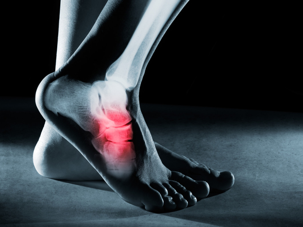
Foot pain can be crippling. Where is Navicular Bone? Why Does my Navicular Bone Hurt? How do you treat Navicular bone pain? Let’s dig in.
Where is Navicular Bone?
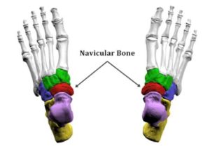
There are 26 bones in the foot. The Navicular Bone is a small C-shaped bone located on the inside portion of the midfoot. It is nestled between the talus, the three cuneiform bones, and is a structural link between the midfoot and forefoot. It provides important support of the foot and arch during movement.
The Tibialis Posterior is the only muscle that attaches to the Navicular Bone and is also important in supporting the arch (1). Ligaments are thick bands of connective tissue that connect one bone to another. The 26 bones in the foot are held together by ligaments and muscles. The spring ligament spans between the heel bone and the Navicular Bone and is an important ligament as it provides support for the arch and foot (2).
Why Does my Navicular Bone Hurt?
There are multiple causes of Navicular Bone pain. The most common include:
Fractures
Fractures typically occur as a result of trauma or chronic overuse. Navicular fractures account for 14-35% of all stress fractures (3) Symptoms include pain, swelling, and some limited range of motion. A CT scan is the study of choice as a high percentage of fractures are missed on plain x-ray (4)
Osteoarthritis
Osteoarthritis involves the progressive wearing down of cartilage. Cartilage protects the joint and enables the joint to move smoothly. The most common causes of Navicular osteoarthritis are degeneration and trauma (4). Pain is aggravated by prolonged standing and walking is common.
Ligament Instability
Ligaments provide stability for the 26 bones in the foot. Ligament injuries can compromise this stability leading to an unstable foot and arch. This instability allows for excessive and abnormal movement of the bones and tendons leading to degeneration, arthritis, and tendonitis.
Low Back Nerve Injury
Low back nerves provide the electrical signal to the muscles in the legs, ankle, and feet. This signal allows for contraction and relaxation of the muscles. Muscle strength is critical to the stability of the foot. Disc bulges, herniation, and slippage can reduce this electrical signal leading to foot instability and pain.
Accessory Navicular Syndrome
The Accessory Navicular is an extra piece of bone attached to the Navicular Bone. It occurs in 4-14% of the population (6). They are typically located on the inside aspect of the Navicular Bone. Accessory Navicular bones are classified into three groups based upon shape and position (7). Accessory Navicular syndrome occurs when the extra piece of bone causes pain. It typically occurs after a trauma or from shoes rubbing against the extra bone. Pain and swelling are common.
Treatment options depend upon the specific cause of the Navicular Bone pain, Treatments are typically divided between nonoperative and operative.
Fractures: Treatment is dependent upon the severity of the fracture, its location, and the presence of any fragments. Immobilization with no weight-bearing is the treatment of choice when possible. Multiple surgery options exist.
Osteoarthritis: Conservative care involves activity modification, weight loss, orthotics, walking boot, and oral medications including fish oil and turmeric. Steroid injections are oftentimes recommended if pain persists. Steroids are powerful anti-inflammatory agents that are toxic to cartilage and should be avoided (8). Surgery options include fusion and joint replacement.
Accessory Navicular Syndrome: Immobilization, safe anti-inflammatory medications, physical therapy, and orthotics. Surgery involves removing the extra piece of bone.
Ligament Instability: Rest, elevation, and immobilization are first-line treatment options. Surgery is necessary for ruptures where both ends of the ligament are physically separated.
Low Back Nerve Injury: Treatment is dependent upon the severity of the specific condition and amount of nerve dysfunction.
Are there regenerative, natural treatment options? Absolutely!
Precise, ultrasound and x-ray guided injections can treat Navicular Bone pain due to arthritis, ligament or tendon injuries, and low back dysfunction. The doctors at Centeno-Schultz Clinic are experts in the treatment of foot, ankle, and leg injuries. Board-certified, fellowship-trained physicians provide multiple treatment options including PRP and the use of your own stem cells. These procedures require extensive training and can not be performed by your Primary Care Physician (PCP) or orthopedist. To watch a PRP injection into the foot, see the video below.
3 Things to Know About Navicular Bone Pain
Common causes of navicular bone pain are fracture and arthritis
Important but less appreciated causes include ligament injury, irritation of low back nerves, and Accessory Navicular Bone.
PRP and stem cells are effective, nonsurgical, natural treatment options for Navicular Bone pain.
In Conclusion
The Navicular Bone is a small C-shaped bone located on the inside portion of the midfoot. It provides important support of the foot and arch during movement. Fracture and arthritis are common causes of pain. Less common but other important causes of Navicular pain include ligament injury, irritation of low back nerves, and Accessory Navicular syndrome. Don’t be sidelined by ongoing foot pain. Schedule a Telemedicine evaluation and learn about your non-surgical, natural treatment options.
4. Thompson FM, Mann RA: Degenerative arthritis of the talonavicular joint. Surgery of the foot and ankle. Edited by: Mann RA, Coughlin MJ. 1992, St. Louis: Mosby, 636-637. 6
7.Wynn M, Brady C, Cola K, Rice-Denning J. Effectiveness of Nonoperative Treatment of the Symptomatic Accessory Navicular in Pediatric Patients. Iowa Orthop J. 2019;39(1):45‐49.
8.Wernecke C, Braun HJ, Dragoo JL. The Effect of Intra-articular Corticosteroids on Articular Cartilage: A Systematic Review. Orthop J Sports Med. 2015;3(5):2325967115581163.
Download your copy!
This groundbreaking work outlines a new way of approaching orthopedic health and treatments, laying down the foundation for Interventional Orthopedics. Download your copy today!