Are you getting an MRI of your thoracic spine? Get the help you need with the Centeno-Schultz Clinic. Learn what you need to know about this exam. What to expect on your thoracic spine MRI scan? What exactly is an MRI of the thoracic spine? How does an MRI work? What are the risks and benefits associated with an MRI? When should you need a thoracic spine MRI? What is the process of doing a thoracic spine MRI? What would an abnormal thoracic spine MRI indicate? Let’s dig in.
What To Expect On Your Thoracic Spine MRI Scan
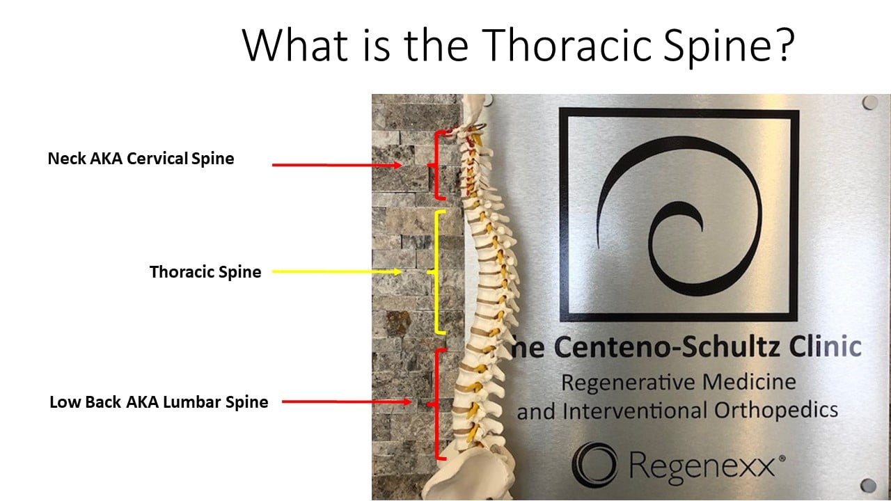
Thoracic spine pain that is unresponsive to conservative care and medications warrants further evaluation. Your doctor may order a thoracic spine MRI, a radiographic study that provides detailed information about the structures in the med back. The thoracic spine is that portion of the spine that is below the neck and above the low back.
What Is An MRI Of The Thoracic Spine?
A thoracic spine MRI scan is a very powerful medical imaging tool that is used to evaluate pain, numbness, and dysfunction in the thoracic spine. It takes very detailed pictures of the spine, joints, muscles, tendons, ligaments, muscles, nerves, and spinal cord in your mid back to identify any abnormalities which may be responsible for your pain.
What To Expect On Your Thoracic Spine MRI Scan
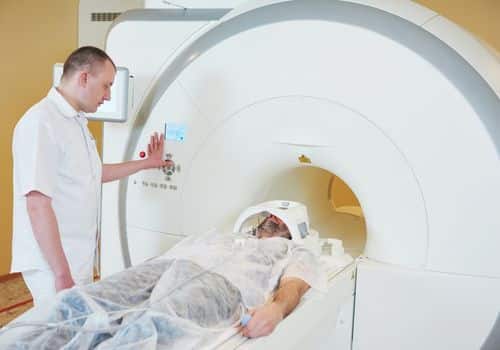
MRI scan of the thoracic spine requires an appointment either at a free-standing facility or in a hospital. NPO and medication status are discussed in detail below. Thoracic spine MRI without contrast takes between 15- 30 minutes. There are two types of MRI scans: Open and Closed. In both, you are laying on your back during the scan. Ear protection is provided as there are loud crackling noises produced by the MRI scanner. MRIs ordered with contrast require that the patient have an IV. This allows the MRI technician to inject the appropriate contrast material.
Most scans are performed with the patient awake and not medicated. Patients that have anxiety or that are claustrophobic may require an oral sedative or IV sedation.
What Is An MRI Of The Thoracic Spine?
MRI is an abbreviation for magnetic resonance study. MRI of the thoracic spine is a type of non-invasive medical imaging that provides three-dimensional detailed images of thoracic regions and their contents.
How Does An MRI Work?
Magnetic resonance imaging (MRI) is a medical imaging technique that uses a magnetic field, radio waves, and a computer to create detailed images of targeted areas in your body (1). Unlike x-ray and CT scans, MRI does not use radiation. Rather it uses a high-power magnet that forces the protons in the body to align with the magnetic field. Radio waves are then applied which stimulate the protons out of alignment. When the radio waves are turned off, the MRI sensors record the energy released as the protons realign with the magnetic field. The energy is then converted into pictures that your doctor uses to assess your condition.
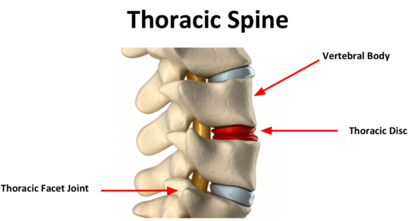
The thoracic spine is composed of 12 boney building blocks called vertebral bodies. Sandwiched between each vertebral body is an important shock absorber referred to as a disc. Housed within the boney protection of the spine is the spinal cord and exiting nerve roots. Ligaments are thick pieces of connective tissue that provide stability.
Benefits And Risks Associated With MRI
There are risks and benefits associated with an MRI of the thoracic spine which include:
Benefits
Noninvasive imaging study that does not involve radiation.
Able to visualize soft tissue areas such as muscles, tendons, ligaments, and discs.
Painless
Risks
No risk to the average patient when safety guidelines are followed
Noisy
The magnetic field may cause implanted medical devices such as pacemakers to malfunction. May also compromise image and study quality.
May cause anxiety in patients with claustrophobia.
Allergic reaction if contrast is used and the patient is allergic.
When Would You Need An MRI Scan?
There are a number of indications for a thoracic spine MRI which include (2):
Thoracic pain that is unresponsive to conservative care and medications.
Suspected thoracic spine infections such as discitis and osteomyelitis.
Suspected thoracic spine fractures after trauma
Abrupt onset of leg weakness and numbness consistent with possible spinal cord compression.
Vein and artery abnormalities
Hemorrhage
Suspected tumors
Unexplained progression of symptoms in patients with scoliosis
Thoracic spine MRI is a valuable scan utilized by your doctors in evaluating your thoracic pain and dysfunction. To learn more about what to expect before, during, and after the MRI please read below.
Before The Procedure
NPO Status: Patients scheduled for a thoracic spine MRI will be asked not to eat or drink typically six hours prior to the study. This is to prevent any stomach contents from entering the lungs in the event you become nauseous or vomit during the procedure.
Medications: Please take all scheduled medications prior to the MRI of the thoracic spine.
Timing: Like most medical appointments, you most likely will be asked to arrive at the MRI facility 15 minutes prior to your appointment time. The MRI facility may be free-standing or a department in the hospital. Intake paperwork will be completed and review past medical history, current medications, presence of indwelling metal such as knee or hip replacement, metal plates or screws, pacemakers, or spinal cord stimulators. If your doctor ordered an MRI with contrast, an IV will be started in either your hand or arm.
During The Procedure
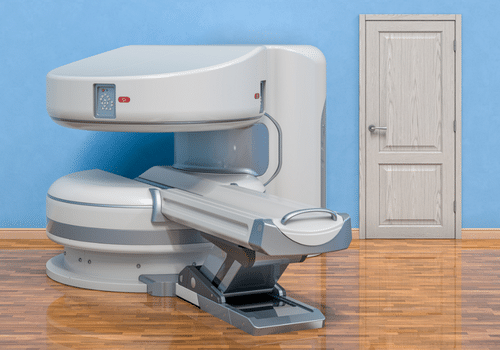
There are two main types of MRI machines: open MRI unit vs closed MRI unit.
A closed MRI machine is cylindrical in shape and the patient is advanced into a tunnel for the study. The closed unit completely envelops the patient during the scan. It can be claustrophobic and cause anxiety. The open MRI on the other hand has a wider opening with magnets on both the top and bottom of the machine.
During the scan, you are asked to lie on your back. Hearing protection is provided as the MRI machine makes a series of loud clicking noises. You will be instructed to stay as still as possible as movement will compromise the images and study. The length of a thoracic spine MRI is 15-30 minutes without contrast.
After The Procedure
Patients that did not receive sedation typically feel no adverse effects from the MRI scan. They can leave immediately after the scan and resume normal daily activities. Patients that receive sedation typically are monitored until they meet discharge criteria. Most feel sleepy for a portion of the day due to the sedation medications.
Who Assesses Your Results?
A radiologist will review your thoracic spine MRI within 24-48 hours and provide a written report. The MRI report is then sent to the referring doctor. Many physicians have digital access to the MRI scan and review the images.
What Would An Abnormal Test Result Indicate?
A thoracic spine MRI is a very detailed 3D examination of your spine, bones, ligaments, tendons, nerves, blood vessels, and spinal cord (3). Abnormalities may be detected. The most common abnormalities include:
Vertebral fractures: a fracture of one or more of the boney building blocks that make up the thoracic spine. This is typically seen in trauma and osteoporosis.
Injuries of the surrounding ligaments: ligaments are the duct tape that connects bone to bone. They are susceptible to sprain, stretching, tearing, and rupture.
Infection: may involve the disc (discitis), the bone ( osteomyelitis), the spinal cord layers (meningitis), or the spinal cord itself.
Spinal cord compression: a significant injury that may be due to a slipped disc, spinal tumor, overgrowth of facets, disc protrusions or herniations, and infection.
Slipped discs: misalignment of the boney building blocks with potentially significant clinical consequences.
Pressing of your nerves or spinal cord: may occur as a result of disc bulge, herniation, facet overgrowth, infection, and ligament overgrowth
Tumors
Unusual curves in the spine: examples include scoliosis or kyphosis.
Get The Best Diagnosis For Your Thoracic Spine Condition
MRI of the thoracic spine is a powerful noninvasive diagnostic tool that provides detailed images of the thoracic spine and its many parts. The Centeno-Schultz Clinic are experts in the evaluation and treatment of thoracic pain. We use the information in the thoracic spine MRI along with findings on ultrasound examination, physical examination, and review of medical history to accurately diagnosis the source of one’s thoracic spine conditions. Why? An accurate diagnosis is required to develop the very best treatment plan. if the diagnosis is incorrect the clinical results often are disappointing leaving the patient in continued pain.
In Conclusion
MRI thoracic spine is an important medical imaging tool used to evaluate pain, numbness, and dysfunction in the thoracic spine. Magnetic resonance imaging (MRI) uses a magnetic field, radio waves, and a computer to create detailed images of the targeted area. Benefits of MRI include no radiation, no discomfort, and the ability to see soft tissues. The risks include potential allergic reactions when contrast is used, anxiety in those that are claustrophobic, and malfunctioning with implanted medical devices. Indications for thoracic spine MRI include pain unresponsive to conservative care, suspected infection, fracture, vascular abnormalities, hemorrhage, and tumors. What to expect before, during, and after the procedure is discussed.
Centeno-Schultz Clinic is here to provide you with all the assistance you need. Download a copy of the Spine Owner’s Manual today.
If you or a loved one suffers from persistent thoracic pain that has not responded to conservative care please schedule a new patient evaluation. For those not in the Denver Boulder area we offer telemedicine appointments. A board-certified, fellowship-trained physician will review your history, symptoms, and radiographic studies and discuss possible regenerative treatment options. To schedule please contact Jen at 720-287-7196 or [email protected] or Vanessa at [email protected].
2.Tanaka Y, Ohno Y, Hanamatsu S, Obama Y, Ueda T, Ikeda H, Iwase A, Fukuba T, Hattori H, Murayama K, Yoshikawa T, Takenaka D, Koyama H, Toyama H. State-of-the-art MR Imaging for Thoracic Diseases. Magn Reson Med Sci. 2022 Mar 1;21(1):212-234. doi: 10.2463/mrms.rev.2020-0184. Epub 2021 Apr 29. PMID: 33952785; PMCID: PMC9199970.
3.van Beek EJR, Kuhl C, Anzai Y, Desmond P, Ehman RL, Gong Q, Gold G, Gulani V, Hall-Craggs M, Leiner T, Lim CCT, Pipe JG, Reeder S, Reinhold C, Smits M, Sodickson DK, Tempany C, Vargas HA, Wang M. Value of MRI in medicine: More than just another test? J Magn Reson Imaging. 2019 Jun;49(7):e14-e25. doi: 10.1002/jmri.26211. Epub 2018 Aug 25. PMID: 30145852; PMCID: PMC7036752.