The headaches and dizziness became progressive in nature and responsive to conservative care. Chiropractic adjustments provided only transient relief as you could not maintain your adjustment. A surgeon reviewed your history, symptoms and radiographic studies. He mentioned different measurements that were abnormal. What is the Atlantodens Interval? What is the Atlas? What is the Axis bone? What is the Atlantoaxial Joint? What stabilizes the Atlantoaxial Joint? What are the unique problems associated with the Atlantoaxial Joint? What is Atlantoaxial Joint Instability? How is the Atlantodens Interval measured? What are the normal Atlantodens Interval measurements? What are the step by step instructions on how to measure the Atlantodens Interval? Let’s dig in.
Introduction
Problems with the head/neck stability can be devastating to both patients and their families. Unfortunately, most clinicians do not understand Craniocervical or Atlantoaxial Instability and often times dismiss patient complaints. Patients are misdiagnosed or labeled are psychologically unbalanced. We are committed to informing patients and providers alike on the importance of their two conditions.
What is the Atlantodens Interval (ADI)
The atlantodens interval is a radiographic measurement that is used in the evaluation of upper cervical spine conditions. It can also be referred to as the atlantodental interval. The atlandodens interval measurement is most commonly used in the evaluation of atlantoaxial instability (AAI).
Relevant Cervical Anatomy
Review of the upper neck anatomy will help with understanding this radiographic measurement and the assoicated clinical conditions. One of the most confusing aspects of cervical anatomy is due to the fact that the uppper neck bones which are numbered also have specific names. Deep breath.
What Is the Atlas? (C1 Bone)
Atlas AKA C1 Bone
The Atlas bone, also known as the C1 is the first bone in your neck (1). It is named after the Greek God Atlas who held up the world on his shoulders. The Atlas plays an important role as your head rests directly on this bone.
What Is the Axis Bone
Axis AKA C2 Bone
The Axis bone is the second bone in the neck and as such it is also referred to as C2 (2). It is an odd-looking bone as it has a boney peg that looks like the Washington Monument. This boney projection is called the Dens. The Dens extends upward to the Atlas bone. It provides a stable point upon which the Atlas bone can rotate.
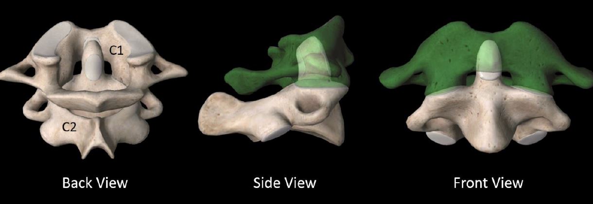
What Is the Atlantoaxial (AA) Joint?
The AA joint is formed by the union of the C1 and C2 bones. Here is what the AA looks like from different angles.
Atlantoaxial Joint Stability (C1/2)
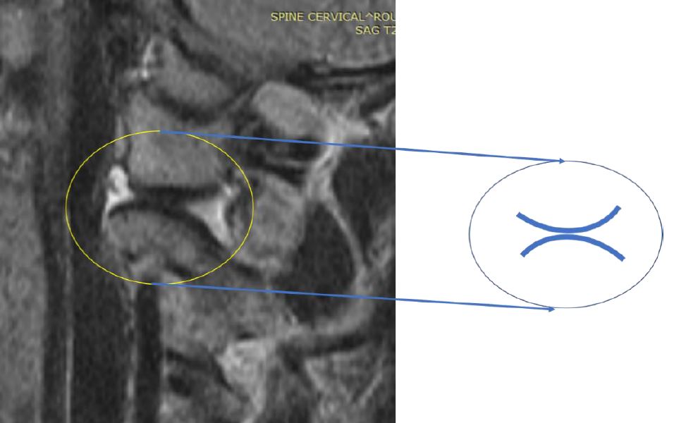
The stability of the Atlantoaxial joint is dependent upon ligaments and muscles of the upper neck. The Transverse ligament is the key stabilizer. It is a thick band that forms a tight seatbelt across the dens. More specifically it arches across the ring of the C1 bone and holds the Dens in tight contact with the C1 bone. It schematically illustrated in cross section below. The blue circle at the top represents the dens AKA C2 bone. The Transverse ligament snuggly holds the Dens in place much like a seatbelt.
Unique Problems with the Atlantoaxial Joint (C1/2 Joint)
The Atlantoaxial joint plays a very important function in the neck as it provides approximately 50% of rotation for the neck (2). Like all joints in the spine it lined with cartilage and has a joint capsule. What is unique and problematic about the AA joint is the shape of its internal joint surfaces. Most joints fit snuggly together as the joint surfaces fit together like a puzzle. It is the classic ball socket configuration. A common example includes the joint hip where the ball of the Femur fits snuggly into a socket. A convex surface fits snuggly into a concave surface as illustrated below. This configuration is stable.
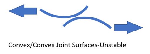
Unfortunately the AA joint does have one joint surface fitting nicely into the other. Why? Because both surfaces of the joint are convex as illustrated below.
The convex/convex configuration makes for an inherently unstable joint as illustrated below.
Atlantoaxial Instability (AAI)
Atlantoaxial Instability is a medical condition where there is excessive movement betwen the C1 and C2 bone (3). The loss of stability of the AA joint can occur as a result traumatic, inflammatory and cogenital abnormalities (4). Examples of congenital conditions include Down’s syndrome and patients with hypermobility such a Ehlers-Danlos Syndrome. To learn more about the Ehlers-Danlos syndrome and Craniocervical Instability please click here.
How Is the Atlantodens Interval Measured?
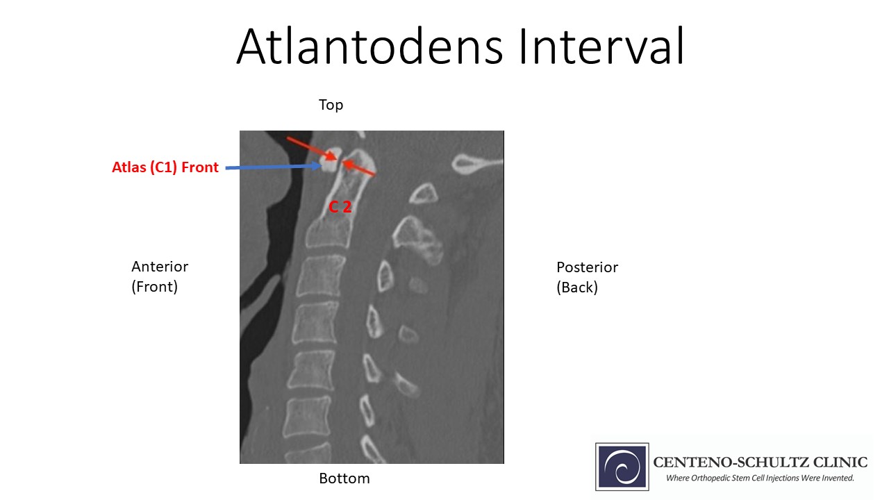
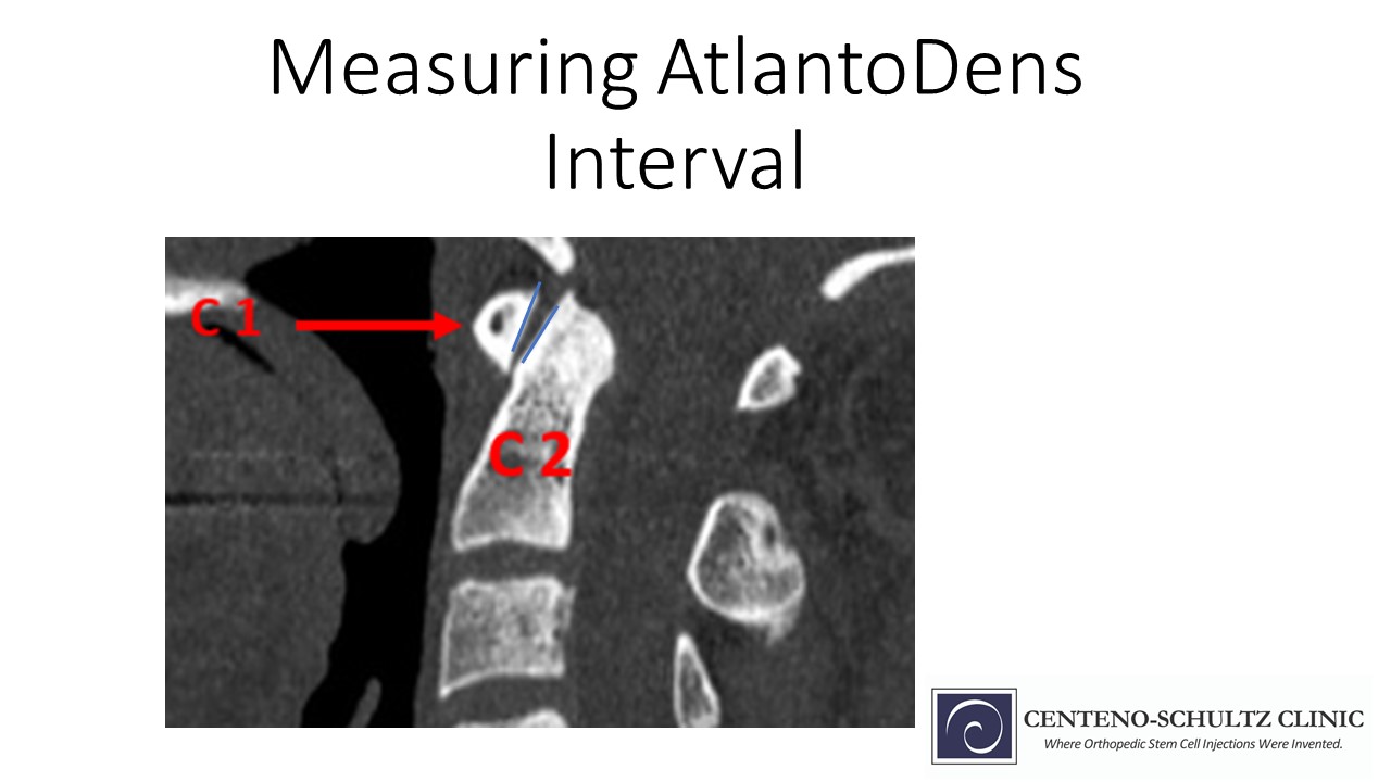
The Atlantodens Interval is the distance between the backside of the C1 bone and the front of the C2 bone. It is measured in milimeters (mm). To accurately assess the presence of Atlantoaxial Instability it is important that the radiographic study includes both flexion and extension images. Why? Traditional radiographic studies are static as they have the patient lying still typically lying flat. Instability is best evaluateed during static tests with the patient upright and flexed forward and extended back.
What Are Normal Atlantodens Interval Measurements?
The Atlantodens Interval can be measured on x-rays, CT and MRI scans. The normal ADI for adults is less than 3 mm whereas it is less than 2mm for children.
Step by Step Instructions on How to Measure the Atlantodens Interval
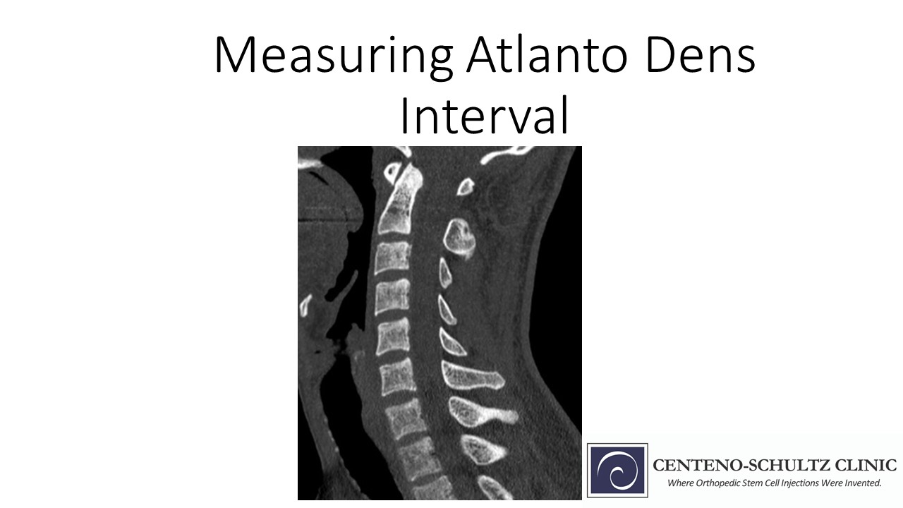
Start with a good quality sagittal CT, x-ray or MRI image of the cervcial spine. AKA sideview
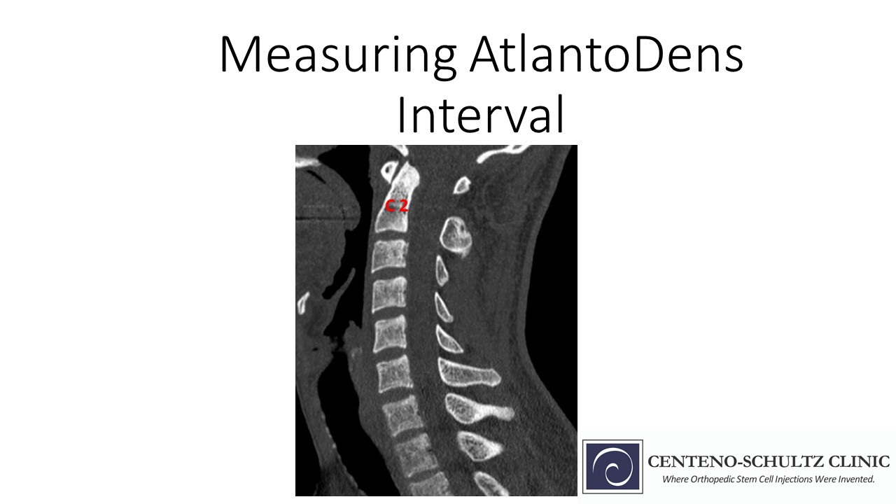
Identify the C2 bone AKA dens
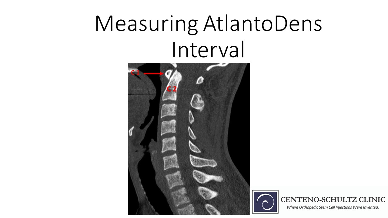
Identify the front of the Atlas AKA C1 bone
4. Identify the backside of the C1 bone and draw a line. Next identify the front aspect of the C2 bone and draw a line.
5. The distance between the backside of the Atlas and the front portion of the C2 is the Atllantodens Interval. It is ilustrated by the double sided arrow. It is measured in mm.
In Summary
The Atlantodens Interval also known as the Atlantodental Interval (ADI) is a radiographic measurement used in the evaluation of upper cervical spine conditions.
The Atlantodens Interval is most commonly used in the evaluation of the Atlantoaxial Instability (AAI).
The Atlas also known as the C1 is bone is the top bone in the neck.
The Axis is the second bone in the neck and is also known as the C2.
The Atlantoaxial joint is the union of the C1 and C2 bone. It’s stability is dependent upon the ligaments and muscles of the upper neck.
The Transverse ligament is a key stabilizer and forms a seatbelt around the C2 bone.
Most joints are classic ball socket configuration. This convex/concave configuration is inherently stable.
The AA joint has a convex/convex configuration and is inherently unstable.
The Atlantodens Interval is the distance between the backside of the C1 bone and the front of the C2 bone. The distance between the two is the ADI and is measured in mm.
At the Centeno-Schultz Clinic we are experts in the managment of upper cervical conditions such as Craniocervical and Atlantocervical Instability. To learn more about Atlantoaxial Instability and Craniocervical instability please download our newest book: CCI 101 by clicking here.
If you or a loved one suffers from persistent headaches, neck pain and brain fog and has not responded to conservative therapy please schedule a New Patient Evaluation. A board certified, fellowship trained physician will compassionately review your history, symptoms and radiographic studies and discuss possible treatment options.
1.Mead LB 2nd, Millhouse PW, Krystal J, Vaccaro AR. C1 fractures: a review of diagnoses, management options, and outcomes. Curr Rev Musculoskelet Med. 2016;9(3):255-262. doi:10.1007/s12178-016-9356-5.
2.Klimo P Jr, Rao G, Brockmeyer D. Congenital anomalies of the cervical spine. Neurosurg Clin N Am. 2007 Jul;18(3):463-78. doi: 10.1016/j.nec.2007.04.005. PMID: 17678749.
3.Yang SY, Boniello AJ, Poorman CE, Chang AL, Wang S, Passias PG. A review of the diagnosis and treatment of atlantoaxial dislocations. Global Spine J. 2014;4(3):197-210. doi:10.1055/s-0034-1376371
4.Subin B, Liu JF, Marshall GJ, Huang HY, Ou JH, Xu GZ. Transoral anterior decompression and fusion of chronic irreducible atlantoaxial dislocation with spinal cord compression. Spine (Phila Pa 1976). 1995 Jun 1;20(11):1233-40. doi: 10.1097/00007632-199506000-00004. PMID: 7660230.