We’re nearing the quarter-century point of the 21st century, and technology in the field of medicine continues to advance at an incredibly fast pace. Thanks to advances in imaging technology, we’ll learn more in the following few decades about the neuromusculoskeletal system than we’ve learned in the past few hundred years. A clear example of this comes from two doctors, Scott Rosa and Dave Harshfield, who have discovered how to measure the flow of cerebrospinal fluid (CSF) around the brain using MRI, which can help patients with CCJ instability. We’ll explore this in a moment, but first, let’s define CSF and CCJ instability.
What Is CSF?
Cerebrospinal fluid (CSF) is a clear fluid that lives in the brain and spinal cord. It functions as both a cooling and waste-removing mechanism and also supplies nourishment to the brain. A variety of diseases, such as Alzheimer’s and multiple sclerosis, have been potentially associated with the poor flow of CSF. On brain imaging, CSF flow has a similar appearance to blood flowing, as CSF, too, is pumped through the brain by the body.
Unfortunately, MRI technology isn’t good at handling movement, so in the past, we haven’t had the ability to observe CSF flow back and forth between the brain to the spinal cord. However, in recent years, Fonar, a company that makes stand-up MRIs, worked with select physicians at developing protocols for CSF flow measurement. In fact, the company has worked with NASA on the disrupted CSF flow in astronauts, which was in the news just a couple of years ago.
What Is CCJ Instability?
Very strong, tough ligaments attach the head to the cervical (upper) spine. When there is a trauma, such as a car crash, that results in some type of blow to the head, these ligaments can be injured, leading to CCJ instability. Symptoms can include headaches, dizziness, disorientation, and so on. We have a novel therapy that can treat these ligaments and CCJ instability, and we’ve had good results. To learn more about this, see the video above.
CSF Flow Can Now Be Seen via MRI
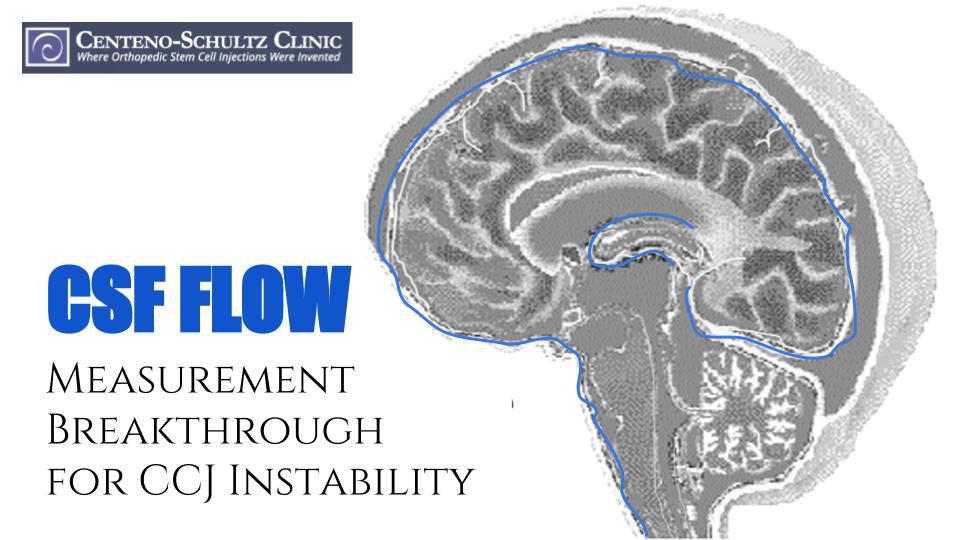
Dr. Centeno has worked with Dr. Rosa and Dr. Harshfield, treating their CCJ instability patients with our novel stem-cell-based injection technique, and has reviewed many images like the amazing one shown above.
The image on the left is before Dr. Rosa performed one patient’s upper-cervical adjustment, and the image on the right is after. The pulsing black, or dark-colored, fluid that’s moving around the brain and spinal cord should be your focus here. The video shows where these changes are and the fact that these occur after the chiropractic care.
Why Is This Technology Important?
There are some major clinical uses for this MRI technology.
First, patients with significant CCJ instability can benefit. How? There is a lot of space around the spinal cord in the upper spine. This means that even significant upper-neck instability may not necessarily compress the structure to the same degree it would in a lower area of the neck where the canal is smaller. However, these images show that just a small amount of motion disturbance in the upper neck can reduce CSF flow. So the result after chiropractic manipulation? Improvement of CSF flow and more normal movement of the neck bones.
Second, when there is cervical stenosis, disturbances in CSF flow can be found using this MRI technology to measure the flow. With stenosis, pinching or compression of the spinal cord can occur due to neck arthritis. Finally, with recent research showing that the development of Alzheimer’s may be related to CSF flow, this technology may become important for prognosis and diagnosis of the disease.
It’s an exciting century to be pioneering and leading the field of interventional orthopedics as these fascinating advances help us discover ways to streamline our diagnostic and nonsurgical treatment methods for CCJ instability and other neuromusculoskeletal issues at the CCJ Instability Institute.