In a previous blog I discussed the clinical success of rotator cuff repair using expanded stem cell therapy.
Today we had the opportunity to review MRI images of an elderly patient who also underwent the Regenexx procedure 2 years ago for a supraspinatus tear. AB is an 80 y/o patient with neck, headache and shouder pain. Her shoulder pain was severe and she was unable to lift her shoulder. She declined surgery and elected to proceed with mesenchymal stem cell therapy. Her own stem cells were injected into the rotator cuff tear under x-ray guidance.
To understand the differences in pre and post MRI’s, some basic MRI concepts and anatomy is essential.

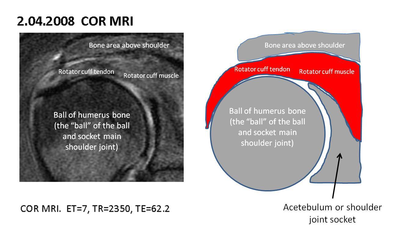
The image above is the patient’s pre-injection coronal MRI. The rotator cuff tendon is the area of interest. The rotator cuff is compromised of 4 principle muscles. Muscles have two parts: the muscle belly and the attachment of the muscle to bone(tendon). Tears in the rotator cuff commonly involve the tendon.

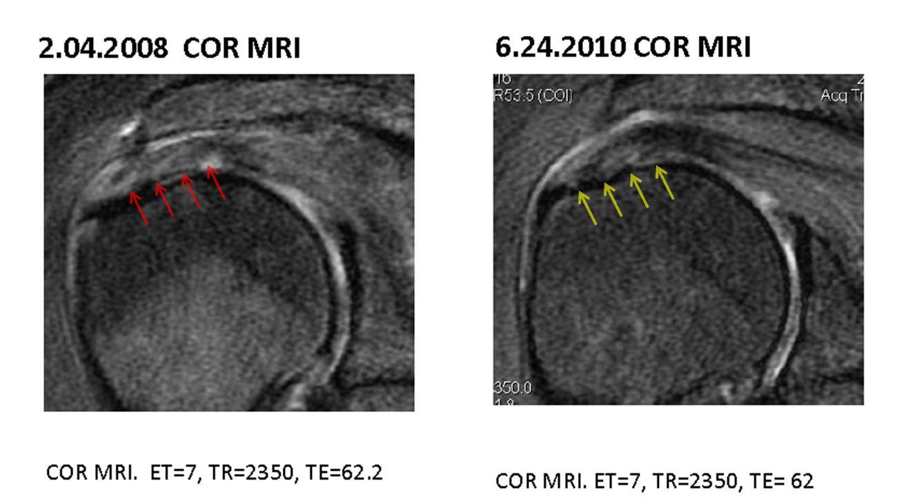
Above are AB’s pre and post MRI’s . On the left the rotator cuff tendon(red arrows) are bright in color and mottled in appearance. This means that it’s a full thickness tear with severe degeneration. On the right is AB’s MRI 2 year post stem cell injection. The rotator cuff tendon identified by the yellow arrows is better organized and darker in color consistent with significant healing. This is consistent with her clinical improvement. She reports 100% improvement in pain and full range of motion.
