Knee Pain
Get Help With Knee PainHaving Knee Pain?
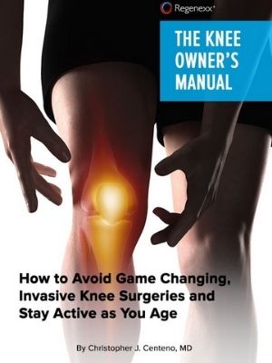
Knee pain can be caused by many factors. Overuse injuries, direct trauma to the knee, and arthritis are the most common causes of knee pain. Damage to the knee structures may cause swelling, scar tissue formation (fibrosis), and loss of function of the joint. Pain is often accompanied by difficulty walking, weakness, and instability.
When the knee is overused, the thigh and shin bones (femur and tibia), cartilage, or tendons may experience stress. This leads to pain and discomfort as well as stiffness in the knee. Overuse injuries are common among athletes who participate in sports that involve running, jumping, or quick turns and stops such as basketball, volleyball, or tennis.
Common injuries and conditions:
- Ligament injuries (sprains and tears), examples include ACL, PCL, MCL and LCL tears
- Meniscus tear – C-shaped fibrocartilage structure between the femur and tibia that functions as cushion. Susceptible to irritation and tears.
- Patellar tendonitis (inflammation of the tendon that attaches the kneecap to the shinbone)
- Hamstring tendinopathy
- Nerve Irritation
- Baker’s cyst (collection of fluid behind the knee)
- Patellar tendon tear – attaches the bottom of the kneecap (patella) to the top of the shinbone (tibia).
- Nonunion Fracture- a bone that has failed to heal after an extended period
- Knee Instability
Conditions Treated At Centeno-Schultz Clinic
ACL Tears
The Anterior Cruciate Ligament (ACL) is one of four major ligaments in the knee. It is an important stabilizer of the knee and prevents the shin bone (tibia) from sliding in front of the thigh bone (femur). The ACL is susceptible to injury. It is most likely to be injured during activity or by impact. A torn ACL is a common injury for athletes at all levels, but it is most common for people who are active or who experience impact injuries to the knee. ACL injuries can happen to anyone of any age, condition, or ability, and it can be injured in many ways. Examples include abruptly changing direction, slowing down while running, landing incorrectly, or getting struck by someone or some object.
Read More About ACL TearsBaker’s Cyst
A Baker’s cyst can cause knee pain. A Baker cyst is swelling caused by fluid from the knee joint protruding to the back of the knee. They are NOT a true cyst since it has communication with the synovial sac. They typically arise from degenerative changes or injury to the articular cartilage (arthritis) or meniscus. At the Centeno-Schultz Clinic, we believe that baker’s Cysts are simply a barometer of the health of the knee joint. In a healthy knee, there are absent whereas with injury and degenerative changes they are common. They arise between the tendons of the medial head…
Read More About Baker’s CystBone Spurs In The Knee
Bone spurs, also known as osteophytes, are abnormal growths that can form along any bony surface in the body. They most frequently occur where tendons and ligaments attach to the bone. As a result, they are more commonly seen in large mobile joints that support weight, such as the hips, spine, ankles, or knees. Surprisingly, most bone spurs are relatively benign but are a clinical sign of instability in the area. Research demonstrates that if a ligament has laxity, the constant strain at the insertion into the bone will elevate the surface of the bone, creating additional bone formation – this is known as a traction osteophyte.
Read More About Bone Spurs In The KneeChondromalacia
Chondromalacia is the knee usually causes pain, typically around the kneecap or deep in the kneecap. You can also have some grinding sensations or crepitus which are sounds and noises coming from around the knee with certain motions. Typically, pain and grinding sensations are worse with bending the knee, especially for prolonged periods of time, kneeling on the knee, walking downstairs, or running downhill. Standing after prolonged sitting or an immobility period where the knee is bent can cause some discomfort as well. Some people may experience swelling, others may experience locking or catching in the knee, feeling the knee wants to give out, or a feeling of weakness….
Read More About ChondromalaciaHamstrings Tendinopathy
Hamstring tendinopathy, a condition that causes pain and tenderness in the hamstring tendons, represents a common but often misunderstood musculoskeletal issue. It typically occurs in athletes and individuals engaged in high-intensity activities but isn’t limited to them. This condition, including its more specific form, high hamstring tendinopathy, can significantly impact daily and athletic performance, making understanding its nuances critical.
Read More About Hamstrings TendinopathyIliotibial Band Syndrome (ITBS)
Also known as “IT Band Syndrome” also known as “ITB Syndrome,” iliotibial band syndrome is a painful medical condition that affects the lateral hip, leg, and knee. It can affect individuals of all ages and most often is caused by repetitive activities like running, cycling, hiking, and walking. Your iliotibial band is a thick band of connective tissue that runs from the outside of your hip down to the outside aspect of your knee. Its principal function is to stabilize the hip and knee. If it becomes tight and dysfunctional, you may experience pain along with this band of tissue due to strain or inflammation. You may also experience pain, limited range of motion in…
Read More About Iliotibial Band Syndrome (ITBS)Knee Arthritis
In the human body, a joint is simply where 2 ends of bone come together. At the ends of these bones, there is a thick substance called “Hyaline Cartilage” that lines the ends. Hyaline cartilage is extremely slippery which allows the two ends of the bone to slide on top of each other. Then there is a capsule that connects the two ends filled with “synovial fluid” that acts as a further lubricant to make it more slippery! Arthritis in the knee is defined by loss of the hyaline cartilage plus other changes that happen to the bone such as additional bone being laid down (bone spurs/osteophytes). The cartilage layer is worn down to the point of exposing the underlying bone they cover…
Read More About Knee ArthritisKnee Instability
Knee instability is a condition that results when the knee joint is unstable and does not move or function normally. This can cause the knee to feel like it is going to give out or buckle. Knee instability can be caused by a variety of factors, including trauma or injury to the knee, ligament injury, arthritis or other degenerative diseases of the knee, weakness or instability of the muscles around the knee, muscle atrophy, injury to another joint in the body creates an imbalance. Knee stability, and stability in general, is very important. Lack of knee stability can lead to more problems over time, such as pain and arthritis…
Read More About Knee InstabilityLCL Sprain
What is an LCL Sprain? A strain or tear to the lateral collateral ligament (LCL) is known as an LCL injury. The LCL is a band of tissue that runs along the outer side of your knee. It aids in keeping the bones together while you walk, ensuring that your knee joint remains stable. How you feel and what type of treatment you’ll require depends on how severely your LCL has been stretched or torn. If it’s only a minor sprain, self-care at home might help. However, if it’s a significant tear or sprain, you may need physical therapy, an injection-based procedure, or surgery….
Read More About LCL SprainLCL Tear
A strain or tear to the lateral collateral ligament (LCL) is known as an LCL injury. The LCL is a band of tissue that runs along the outer side of your knee. It aids in keeping the bones together while you walk, ensuring that your knee joint remains stable. How you feel and what type of treatment you’ll require depends on how severely your LCL has been stretched or torn. If it’s only a minor sprain, self-care at home might help. However, if it’s a significant tear, you may need physical therapy, an injection-based procedure, or surgery. Orthopedists categorize LCL tears into 3 grades…
Read More About LCL TearMCL Sprain
The medial collateral ligament AKA MCL is a thick, strong band of connective tissue on the inside portion of your knee. It connects the top part of the tibia (shin) to the bottom part of the femur (thigh). This is a vital ligament that works along the lateral collateral ligament (LCL), anterior cruciate ligament (ACL), and posterior cruciate ligament (PCL) to bring stability, structure, and movement to the knee. The MCL provides support and stability for the inside (medial) aspect of the knee. MCL sprains are a common injury in sports such as football, hockey, and skiing. The ligament can…
Read More About MCL SprainMCL tear
The medial collateral ligament AKA MCL is a thick, strong band of connective tissue on the inside portion of your knee. It connects the top part of the tibia (shin) to the bottom part of the femur (thigh). This is a vital ligament that works along the lateral collateral ligament (LCL), anterior cruciate ligament (ACL), and posterior cruciate ligament (PCL) to bring stability, structure, and movement to the knee. The MCL provides support and stability for the inside (medial) aspect of the knee. MCL tears are a common injury in sports such as football, hockey, and skiing. The ligament can…
Read More About MCL tearMeniscus Tears
The meniscus is a c-shaped piece of cartilage in the knee that functions as an important shock absorber. It is sandwiched between the thigh and shin bone. There are two menisci per knee. One on the inside portion of the knee (medial) one on the outside aspect (lateral). The knee meniscus is susceptible to injury. The most common injury is a tear in the meniscus. Not all meniscus tears however cause pain. When symptomatic a meniscus tear can cause pain, swelling, and restriction in range of motion. Tears in the knee meniscus can arise from trauma or degeneration. There are many different types of meniscus tears based upon locations….
Read More About Meniscus TearsPatellar Tendon Tear
The Patellar tendon is the thick connective tissue that starts at the base of the kneecap (Patella) and extends down to the shin. The is an extension of the Quadriceps tendon (1). The Quadricep is the large thick muscle that is often referred to as our thigh. The Patellar tendon works together with the Quadriceps muscle to straighten (extend) the knee. It is easy to touch your Patellar tendon as it is immediately below the knee cap. There are many different causes of Patellar tendon tears. Patellar tendon tears are a common sport injury but can also occur from overuse or a motor vehicle injury…
Read More About Patellar Tendon TearPatellar Tendonitis
What is the Patellar Tendon? A tendon is a piece of connective tissue that connects muscle to bone. It serves to move the bone or a given joint. The patellar tendon is a major tendon in the knee. It is located at the bottom of the kneecap (patella) and stretches down to the shin. The patellar tendon enables you to extend your knee, kick, run, and jump. What is Patellar Tendinitis? Patellar tendinitis is an irritation and inflammation of the tendon that connects your kneecap (patella) to your shinbone. Patellar tendinitis, also known as jumper’s knee, can affect anyone. The most common symptom is pain at the shin or lowest part of the kneecap…
Read More About Patellar TendonitisPatellofemoral Pain Syndrome
Patellofemoral pain syndrome (PFS), also called runner’s knee or retropatellar pain syndrome, is a significant cause of pain in the front of the knee. The pain is usually experienced behind or around the patella (kneecap) when the knee is bent or fully loaded. This post discusses everything you need to know about patellofemoral pain syndrome.
Read More About Patellofemoral Pain SyndromePCL Sprain
The Posterior Cruciate Ligament is one of the paired ligaments in the middle of the knee. It is made up of 2 separate bundles: The two bundles of the PCL, and the ALB (anterior lateral bundle), and the PMB (posterior medial bundle), function synergistically to provide stability. The PCL functions as one of the main stabilizers of the knee joint and serves primarily to resist excessive posterior translation of the tibia relative to the femur. The PCL also acts as a secondary stabilizer of the knee preventing excessive rotation specifically between 90° and 120° of knee flexion. A PCL sprain happens when force is applied beyond…
Read More About PCL SprainPCL Tear
The Posterior Cruciate Ligament (PCL) is a paired ligament in the middle of the knee. It is made up of two separate bundles: ALB (anterior lateral bundle) and PMB (posterior medial bundle). These bundles work synergistically to provide stability. The PCL plays an important stabilizing role in the knee joint by resisting excessive posterior translation of the tibia relative to the femur. Between 90 and 120 degrees of knee flexion, it serves as secondary support for preventing excessive rotation. PCL tears happen when force is applied beyond what the PCL tensile strength is capable of resisting. The tensile strength of the PCL is well documented…
Read More About PCL TearPes Anserine Bursitis
Knee pain located at the lower inside of the knee can be caused by Pes Anserine Bursitis, which is irritation of the tendons that run on the inside aspect of the knee. Commonly mistaken for arthritic pain, meniscal pain, and sometimes nerve pain from the low back! Don’t be misdiagnosed, and let’s dive in below to get a better understanding of Pes Anserine Bursitis. The Pes Anserine Bursa is a bursa that surrounds 3 tendons of the leg. A bursa is a thin, slippery, sac-like film that contains a small amount of fluid. A bursa is found between bones and soft tissues in and around joints…
Read More About Pes Anserine BursitisRadial Meniscus Tears
The meniscus is an important fibro-cartilage structure within the knee that absorbs shock and provides cushioning. It has a semicircular ‘C’ shape and sits between the femur (thigh bone) and the tibia (lower leg bone). The meniscus protects the two bony structures from weight, shock, and shear forces. Each knee contains two menisci, one on the outside (lateral) and one inside (medial).
Read More About Radial Meniscus Tears