Knee Hyperextension
Get Help With Knee HyperextensionThere are six different types of joints in the body.
- Saddle joint (i.e. Thumb)
- Gliding Joint (i.e. Wrist)
- Pivot joint (i.e. Forearm)
- Condyloid joint (i.e. Jaw)
- Ball and Socket joint (i.e. Shoulder)
- Hinge Joint
- The hinge joint is like a door, opening and closing in one direction, along one plane. Examples include your elbow joint and your knee joint.
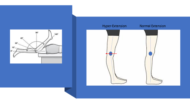
The hinge joint of the knee is designed to open and close from 0 degrees (full extension) to 140 degrees (full flexion). Knee hyperextension occurs when the knee is forced beyond the normal range of motion beyond zero degrees.
What Happens To A Hyperextended Knee?
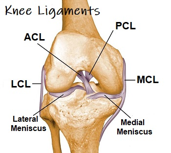
As discussed above, the knee is a hinge joint between the femur and the tibia along with an accessory bone, the fibula. These bones are held together via ligaments that keep the bones aligned while the joint goes through its natural range of motion. These ligaments are:
- Medial Collateral Ligament (MCL): is found on the inner aspect of the knee connecting the femur to the tibia
- Lateral Collateral Ligament (LCL): is found on the outer aspect of the knee and connects the femur to the fibular hear
- Anterior Cruciate Ligament (ACL): is found inside of the knee, connecting the femur to the tibia.
- Posterior Cruciate Ligament (PCL): is found inside the knee, connecting the femur to the tibia in opposite position compared to the neighboring ACL
These ligaments together provide the knee’s overall stability. When working properly the knee motion is from zero degrees fully extended to 140 degrees fully flexed. BUT when a force from the front to the back overcomes the strength of the ligaments the knee is forced into an unnatural position called “Hyperextension” which, in turn, can further damage not only the ligaments but other structures in the joint like the meniscus, cartilage, and bone.
Hyperextension of the knee can occur to anyone, but it’s more common among athletes, especially those who play contact and cutting sports like football, soccer, skiing, or lacrosse. Injuries happen as a result of a direct blow to the knee or forces generated during a quick deceleration or stop (non-contact injury). Multiple factors contribute to the fact that female athletes have a greater risk of knee injury than men, especially those who participate in high-risk sports (1).
What is the difference between a hyperextended knee and hyperextension injury?
Some individuals naturally have ligaments that are “more stretchy” than others which allow the knee to naturally go into knee hyperextension. These individuals walk/stand with their knees in hyperextension and live their entire life without incident while others with normal ligaments get injuries where the knee is forced into hyperextension, beyond the natural limits of their ligaments and result in injury to the ligaments and other structures of the knee!
What Are The Symptoms?
When you get a knee hyperextension injury, there are many signs and symptoms that develop in the knee. When these signs exist, it is recommended to have a qualified physician examine the knee to determine what is damaged, and the extent of the damage. See below as I go through the main signs and symptoms of having knee hyperextension.
Knee Instability
Normal activity such as walking or climbing stairs creates a sense of instability or the feeling that the knee is not going to support your weight and potentially cause a fall.
Feeling The Pain After Injury
Knee pain after an injury is sometimes immediate but often it is a few hours to days later. Damage to the soft tissue structures swells inside the ligaments. The distended ligaments get further stressed with light activity and cause localized pain.
Having Limited Mobility
Swelling in the soft tissue in the knee and surrounding the knee inhibits the ligaments from working properly and limits the range of motion. Swelling inside the knee joint can also build up, also limiting the range of motion.
Water On The Knee
When soft tissue in the knee gets damaged it will swell and leak into the knee joint creating a knee effusion (water on the knee). The knee then becomes puffy above the kneecap and can limit its range of motion.
Spotting Bruises
When ligaments/tendons become damaged, some blood vessels break and cause local bleeding that turns into bruises.
What Are Its Primary Causes?
Knee hyperextension injuries typically occur in a sporting activity that involves contact, jumping, or cutting sports (2). Forceful knee hyperextension via contact or non-contact injuries commonly injure the ACL, PCL (rare), and other structures such as meniscus, and can even cause bone contusion in the femoral condyle or tibial plateau. The extent of the damage can be as little as a small sprain or strain but other high force injuries can cause complete tears in the ligaments.
ACL Tears
The Anterior Cruciate Ligament (ACL) is one of four major ligaments in the knee. It is an important stabilizer of the knee and prevents the shin bone (tibia) from sliding in front of the thigh bone (femur). The ACL is susceptible to injury. It is most likely to be injured during activity or by impact. A torn ACL is a common injury for athletes at all levels, but it is most common for people who are active or who experience impact injuries to the knee. ACL injuries can happen to anyone of any age, condition, or ability, and it can be injured in many ways. Examples include abruptly changing direction, slowing down while running, landing incorrectly, or getting struck by someone or some object.
Read More About ACL TearsEDS in Children
Ehlers-Danlos Syndrome (EDS) refers to a group of disorders that affect the body’s connective tissue including skin, tendons, and ligaments. It is a hereditary disorder which means you are born with it. EDS has many different signs and symptoms which can vary significantly from patient to patient. It most commonly affects the skin, joints, and blood vessels. The estimated prevalence for all EDS varies between 1/10,000 and 1/25,000. The three most common types of EDS are: Hypermobile, Classic, and Vascular. We have used these skills and knowledge to treat the loose ligaments commonly found in EDS in children. Treatment options include bone marrow concentrate (BMC) and PRP.
Read More About EDS in ChildrenEhlers-Danlos Syndrome (EDS)
Disorders that affect and weaken the connective tissues such as tendons and ligaments. It is a hereditary disorder which means you are born with it. EDS has many different signs and symptoms which can vary significantly depending upon the type of EDS and its severity. It most commonly affects the skin, joints, and blood vessels. Joints are typically hypermobile with excessive joint range of motion because of a defect in collagen formation. In most cases Ehlers-Danlos syndrome is inherited. That is to say that you are born with it. The two main ways EDS is inherited are: autosomal dominant inheritance and autosomal recessive inheritance…
Read More About Ehlers-Danlos Syndrome (EDS)Knee Instability
Knee instability is a condition that results when the knee joint is unstable and does not move or function normally. This can cause the knee to feel like it is going to give out or buckle. Knee instability can be caused by a variety of factors, including trauma or injury to the knee, ligament injury, arthritis or other degenerative diseases of the knee, weakness or instability of the muscles around the knee, muscle atrophy, injury to another joint in the body creates an imbalance. Knee stability, and stability in general, is very important. Lack of knee stability can lead to more problems over time, such as pain and arthritis…
Read More About Knee InstabilityPCL Sprain
The Posterior Cruciate Ligament is one of the paired ligaments in the middle of the knee. It is made up of 2 separate bundles: The two bundles of the PCL, and the ALB (anterior lateral bundle), and the PMB (posterior medial bundle), function synergistically to provide stability. The PCL functions as one of the main stabilizers of the knee joint and serves primarily to resist excessive posterior translation of the tibia relative to the femur. The PCL also acts as a secondary stabilizer of the knee preventing excessive rotation specifically between 90° and 120° of knee flexion. A PCL sprain happens when force is applied beyond…
Read More About PCL SprainPCL Tear
The Posterior Cruciate Ligament (PCL) is a paired ligament in the middle of the knee. It is made up of two separate bundles: ALB (anterior lateral bundle) and PMB (posterior medial bundle). These bundles work synergistically to provide stability. The PCL plays an important stabilizing role in the knee joint by resisting excessive posterior translation of the tibia relative to the femur. Between 90 and 120 degrees of knee flexion, it serves as secondary support for preventing excessive rotation. PCL tears happen when force is applied beyond what the PCL tensile strength is capable of resisting. The tensile strength of the PCL is well documented…
Read More About PCL TearDiagnosing The Knee That Is Hyperextended
Most of the time, a musculoskeletal physician such as a sports medicine specialist, an orthopedic surgeon, or a physiatrist can evaluate the knee and should use a diagnostic ultrasound with which they can classify the injuries. After the evaluation, if intra-articular damage is supected, they can send you for an MRI on the knee!
BUT, there are some simple things you can do on your own to see if you need to see a specialist to further investigate. Consider the following tests.
- Weight-bearing – If you can stand on a single leg then that is a good sign, but if you are unable to weight bear on that leg, then potentially a bone bruise or ligament damage causing substantial instability can be present – so you should see a specialist.
- Tenderness to palpation – Compress the tissue around the knee and if you are not able to tolerate deep palpation without significant pain, the potential for high-grade injuries to the ligaments/tendons surrounding the knee exists.
- Swelling / bruising – Inspect the top of the knee cap with the leg extended and if you are able to make out the quadriceps muscle and knee cap then significant swelling in the joint is unlikely, but if the contour is gone and puffy (compared to the uninjured leg) then it’s recommend to have a physician check it out!
What Are The Treatment Options?
At-Home Care
Initially, after knee hyperextension injuries, it is best to get off the leg to allow swelling to reduce and return to normal mechanics. Normal healing time depends on the extent of the injury, but the recovery time can take 4-6 weeks to 4-6 months.
RICE
For acute care immediately after injury, many trainers/therapists/physicians recommend “RICE” – Rest, Ice, Compression, and Elevation.
- Rest– keeping weight off the joint and sometimes using crutches can be of benefit for the short term.
- Ice – ice massage can help reduce the swelling as can alternating ice and heat.
- Compression – ace wraps or knee sleeves can help compress the joint which can help overall stability allowing proper biomechanics
- Elevation – can help the swelling to clear out of the joint faster.
It is important to not take excessive medications that block inflammation such as Ibuprofen, Aleve, or other NSAIDs. These medications, along with high doses of corticosteroids, can actually delay healing and make the condition worse in the long run.
Other natural supplementation such as turmeric, high doses of fish oil in combination with acetaminophen (Tylenol) can help your body heal faster on its own. To read more about proper nutrition, read Dr. Pitt’s Nutrition 2.0 for more!
Physical Therapy & Exercise
After the initial swelling reduces and more normal movement and appearance returns, then seeing a qualified physical therapist can be beneficial in “neuro-muscular re-education” of the joint. With injuries, your body will develop bad habits as a work-around and can lead to further injuries and improper biomechanics. Working with PT can help re-educate back to normal movement patterns as well utilize other modalities such as dry needling, blood flow restriction therapy, and others to further assist in your body’s natural healing.
Surgery
Surgery should be the last option, given ACL reconstruction when compared to conservative therapy has not been seen to be beneficial (3). ACL tears do create chronic instability in the knee that can place additional stress on the knee that leads to osteoarthritis and meniscus tears. But recent research has shown that surgery creates a second hit syndrome that potentially increases the degeneration of the knee and places the knee at higher risk for future knee replacement surgery in recent studies. To learn more, check out Dr. Centeno’s blog on the subject.
Surgery Alternative Treatment
At Centeno-Schultz Clinic, we created alternatives to surgery that use your body’s own regenerative capabilities! Since 2012, we have been using reparative cells found in bone marrow concentrate, injected precisely under live X-ray guidance. In the last decade, we have learned that potentially 70+% of ACL tears can be treated and healed without surgery using this innovative technique that we developed!
Consider Taking A Break To Recover & Rehabilitate
Proper rehabilitation is needed with any injury to allow the body to heal as well as to take a step backward to build core strength in the leg so that when you do recover you can come back stronger and prevent any future injuries!
Strengthening Your Knee Ligaments Through Exercise
If you play any high-risk sports and especially if you are female, then preparing and having an ACL prevention program to strengthen the knees through exercise is critical. Here is the best ACL prevention program that we know of: https://la84.org/a-practical-guide-to-the-pep-program/
Seek Your Doctor’s Help To Recover Swiftly
Knee Hyperextension injuries can be devastating to athletes and can require months and months of rehabilitation. If you have a knee hyperextension injury and are looking to get back faster and stronger then reach out to us and see if you are a candidate for our advanced interventional orthopedic techniques to treat the injury and do not forget a good rehab program as well. Learn more about our advanced, image-guided knee injection procedures today!
References
- Hägglund M, Waldén M. Risk factors for acute knee injury in female youth football. Knee Surg Sports Traumatol Arthrosc. 2016 Mar;24(3):737-46. doi: 10.1007/s00167-015-3922-z. Epub 2015 Dec 24. PMID: 26704794. https://pubmed.ncbi.nlm.nih.gov/26704794/
- Fornalski S, McGarry MH, Csintalan RP, Fithian DC, Lee TQ. Biomechanical and anatomical assessment after knee hyperextension injury. Am J Sports Med. 2008 Jan;36(1):80-4. doi: 10.1177/0363546507308189. Epub 2007 Oct 11. PMID: 17932409. https://pubmed.ncbi.nlm.nih.gov/17932409/
- Wasmaier J, Kubik-Huch R, Pfirrmann C, Grehn H, Bieg C, Eid K. Proximal anterior cruciate ligament tears: the healing response technique versus conservative treatment. J Knee Surg. 2013 Aug;26(4):263-71. doi: 10.1055/s-0032-1329720. Epub 2012 Dec 20. PMID: 23258320. https://pubmed.ncbi.nlm.nih.gov/23258320/