Thoracic Facet Joint Pain – Possible Causes And Treatment Options
Get Help With Thoracic Facet Joint PainThe facet joint is an integral part of the spine. It is a small, paired joint that occurs at each level of the spine. The facet joint limits excessive motion in the spine and provides important spinal stability.
The prevalence of thoracic facet pain in patients with ongoing thoracic pain has been demonstrated to be as high as 42%.(1)
Where Is The Thoracic Facet Joint Located?
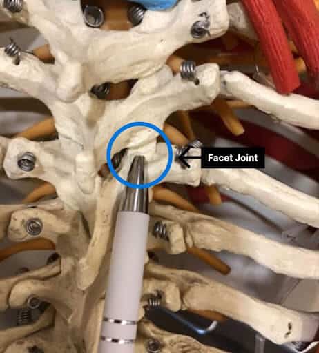
A facet joint is a small paired joint that occurs at each level of the spine. It is located on the backside of the spine immediately adjacent to a large, midline, protruding bone called the spinous process. There is a facet joint on the right and the left of each of the cervical, thoracic and lumbar sections of the spine.
Like all joints, a facet joint is lined with cartilage which is a smooth, white flexible connective tissue that allows the bones to slide smoothly across one another. The joint is surrounded by a cellophane type cover called a capsule which provides stability for the joint.
Both cartilage and the capsule are susceptible to injury which can lead to pain, inflammation, stiffness, and restriction in range of motion.
What Does Thoracic Joint Pain Feel Like?
Symptoms of a thoracic facet joint injury will vary depending upon severity of the injury and which facet joint is injured. The joints have established pain referral patterns (2). Drefus et al demonstrated that pain from a given facet joint does not occur in the immediate area of the joint in 75% of cases.
Rather, it refers to an area away from the joint. For example, pain from injury of the T3/4 facet is felt along the inside border of the scapula. Unfortunately, there is significant overlap between the thoracic referral patterns which can complicate identifying the exact facet joint that is causing the pain.
The most common symptoms associated with a thoracic facet injury are:
- Pain In The Mid-Back: It can be mild, moderate, or severe. Onset can be abrupt or insidious. The pain may be limited to only one side or can occur on both sides.
- Restriction in Range of Motion: Thoracic facet joint injury can damage the joint cartilage making small movements painful.
- Muscle Spasm: Injury to a thoracic facet can trigger significant muscle spasms, leading to pain and limited movement.
- Swelling and Tenderness: Thoracic facet injuries can lead to swelling within the joint and adjacent muscle.
- Radiating pain: If severe, a thoracic joint injury can irritate one or more exiting nerve roots leading to pain wrapping around the chest, into the abdomen, or causing numbness and tingling in the extremities.
What Triggers Or Worsens The Pain?
Thoracic facet pain can be aggravated by any of the following:
- Poor Posture: prolonged computer or screen time can compromise neutral spinal alignment which can aggravate thoracic facet injuries.
- Acute Injuries Or Trauma: trauma can damage both the facet joint capsule and cartilage lining leading to pain, restriction in range of motion and dysfunction.
- Physical Activities: bending, twisting and lifting heavy objects can aggravate thoracic facet joint injuries and pain.
- Repetitive Activities: Engaging in repetitive activities that involve thoracic spinal movements, such as bending, twisting, or lifting, can strain the facet joints and worsen pain over time.
- Coughing
Conditions Possibly Contributing To Thoracic Facet Pain
- Acute Trauma: Motor vehicle accidents and athletic injuries can injure the thoracic facet joint cartilage and capsule leading to pain, swelling, onset of arthritis, and restriction in range of motion. Smith et al documented the presence of blood in the facet joint of patients who died as a result of a motor vehicle injury.
- Postural Syndrome: Neutral spinal alignment allows for optimal spine health and function. Poor posture can place excessive forces on thoracic facet joint capsules and joints leading to injury and dysfunction.
- Ligamental laxity: Ligaments are thick, connective tissue that connect bone to bone. They provide stability and when injured or stretched can lead to varying degrees of instability. Instability, in turn, can lead to facet joint injury and pain.
- Thoracic Spine Subluxation: The vertebral bodies are the boney building blocks of the spine. They stack one upon another in a symmetrical manner. Anterolisthesis is when one vertebral body slides forward in relationship to the others. Retrolisthesis is when a vertebral body slides backwards. Both anterolisthesis and retrolisthesis can injure the facet joint leading to thoracic facet pain.
- Scoliosis: Abnormal sideways curve in the spine can put significant pressure on the affected thoracic facet joints leading to injury, inflammation, and thoracic facet pain.
- Disc degeneration. Sandwiched between each vertebral body is an important shock absorber referred to as a disc. The height and integrity of the disc is important for overall spine function and stability. Disc degeneration can lead to reduction in the height and function of the thoracic discs which in turn can then place excessive pressure and forces onto the facet joints. This excessive pressure can lead to facet joint injury and pain.
- Degenerative Or Arthritic Changes: Poor ergonomics, repetitive activities, and trauma can lead to degenerative and or arthritic changes in the facet joints.
- Fractures: The two bones that make up the facet joint are susceptible to injury and fracture. Fracture can injure both the joint capsule and cartilage leading to thoracic facet joint pain. Cervical facet fractures are relatively common and compromise 10% of all cervical spine fractures (3).
- Tumors: Tumors can occur in the thoracic facet joint, although it is rare. Tumors can be classified as either primary or secondary. The most common primary tumor is a synovial cyst. Secondary tumors occur as a result of metastasis from primary cancers elsewhere in the body.
- Infections: Facet joint infections are rare. It occurs when bacteria, viruses, fungi, or other microorganisms invade the joint. Pain is the most common complaint. It can lead to infectious arthritis also referred to as septic arthritis which is a serious condition. Treatment can include intravenous antibiotics and drainage of the infected joint fluid(4).
When Should You See Your Doctor
Patients experiencing the following symptoms should consider consulting a physician:
Persistent Mid Back Pain: Thoracic pain that is unresponsive to conservative care warrants medical attention. Persistent or worsening mid back pain, either unilateral or bilateral, may be a sign of a thoracic facet joint problem.
Persistent or Permanent Restriction in Range of Motion: Progressive reduction in flexion, extension and rotation of the spine may indicate a thoracic facet injury.
Persistent Mid-Back Muscle Spasms: Everyone has some intermittent, transient muscle tightness. If, however, the mid-back muscle spasms become constant it may be the result of a thoracic facet injury and should be evaluated.
Persistent or Problematic Radiating Pain Across Chest Wall or Abdomen:Thoracic facet injuries can irritate or compress existing nerve roots causing radiating pain across the chest wall or into the abdomen. If radiating pain persists and affects your daily living, it warrants an evaluation.
Numbness, tingling and weakness: Exiting nerves in the thoracic spine can be irritated or compressed by thoracic facet injuries leading to numbness, tingling and weakness in the back, chest or limbs.
Proper Diagnosis Of Thoracic Facet Pain
Diagnosing thoracic facet pain involves a comprehensive evaluation which may include:

- Medical History: Detailed review of current symptoms, onset of pain, aggravating and alleviating factors and treatment to date. Past medical and surgical histories are important as well as current medications and allergies.
- Physical Examination: A thorough physical examination is important and will include range of motion, muscle strength, areas of tenderness, and a neurologic examination.
- X-ray: A nonspecific radiographic test that evaluates bones and is helpful to identify bone fracture or misalignment.
- Magnetic Resonance Imaging (MRI): The preferred imaging choice given its ability to delineate soft tissue. MRI can demonstrate excessive fluid in the facet joint, loss of cartilage, overgrowth and facet cysts(6).
- Computed Tomography (CT): More sensitive than plain x-rays at identifying facet joint injuries (5).
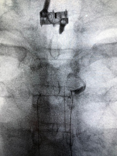
- Diagnostic Thoracic Blocks (Injections): Diagnostic thoracic injections can confirm the diagnosis of a thoracic facet injury. There are two different types of diagnostic injections– intra-articular facet injection and medial branch blocks.
An intra-articular facet injection involves the injection of local anesthetic and steroid directly into the joint under x-ray guidance. The medial branch nerve transmits the pain signal from the injured facet joint to the brain. A medial branch block involves the injection of local anesthetic and steroid along the path of the medial branch nerve.
If there is a reduction in the thoracic pain following either type of injection, the test is referred to as positive and a facet joint injury is likely. A positive intra-articular joint injection together with a positive medial branch block confirms the diagnosis of facet joint injury.
Conventional Treatment Options
Medications: When rest and conservative care fail, medications are often utilized. Common medications include:
- Muscle Relaxants: Can control muscle spasm and restriction in range of motion.
- NSAIDs: Examples include Ibuprofen and Naproxen which are oral agents that can reduce inflammation, swelling and pain. They should be avoided given their significant list of adverse side effects.
- Oral Steroids: Prednisone also known as “ dose pack” are oral steroids that are very powerful anti-inflammatory agents that can reduce inflammation, swelling and pain. These medications should be avoided as they have significant side effects ( link).
- Oral Narcotics: Narcotics such as oxycodone and hydrocodone have powerful analgesics that reduce pain. They have significant side effects which include addiction, tolerance and dependence.
- Conservative Care: When appropriate conservative care should always be first line treatment of thoracic pain. Examples include rest, stretching, massage, acupuncture and application of heat and ice. The goal is to reduce the pain by allowing the body to heal itself.
- Physical Therapy: There are multiple advantages to physical therapy which include:
- Improve strength, flexibility, and stability of the spine.
- Optimize posture and body mechanics
- Manual therapy techniques such as joint mobilization and manipulation that can reduce pain and muscle spasm and restore normal movement patterns.
- Chiropractic Therapy: Multiple chiropractic treatment options can reduce pain and improve function. Examples include spinal adjustments, joint mobilizations, soft tissue therapies, and optimizing neutral spinal alignment and posture
- Spine Injections: When conservative care and medications fail to provide significant benefit, patients are often referred for spinal injections. The three most common injections for the treatment of thoracic facet pain include:
- Intra-Articular Facet Injections: Under x-ray guidance, local anesthetic and steroid are injected directly into the joint. The injection can provide reduction in pain and swelling.
- Medial Branch Blocks (MBB): The medial branch is a nerve that transmits the pain signal from the injured or damaged facet to the brain. During a medial branch block, local anesthetic and steroids are injected along the medial branch nerve, temporarily disrupting the pain signal from being transmitted and registered in the brain.
If the pain is arising from the facet, a medial branch block will provide significant, short term reduction in pain. - Radiofrequency Ablation: Also known as radiofrequency neurotomy or RFA, this is a minimally invasive procedure that burns the medial branch nerve through thermal energy.
A special needle is placed under x-ray guidance along the path of the medial branch nerve. The end of the needle is heated to between 70-90 degrees Celsius (158-194 degrees Fahrenheit). Recall that water boils at 212 degrees Fahrenheit.
The medial branch nerve and the adjacent tissue is cauterized. RFA is a destructive procedure that cauterizes the pain signal from the injured facet joint to the brain.
RFA does not address the underlying facet injury which can progress in nature leading to significant arthritis, pain and restriction in range of motion.
Conditions Associated with Facet Joint Pain in The Thoracic Region
Bone Spurs
Bone spurs, also known as osteophytes, are bony projections that develop along the edges of bones. They are most commonly found in joints — where bones meet — but can also appear on bones where tendons, ligaments, and muscles attach. Bone spurs are often associated with aging and are a common condition. While they can occur in any bone, they’re most often found in areas such as the spine, shoulders, hands, hips, knees, and feet. Bone spurs form as the result of the body trying to repair itself by building extra bone. This process can be triggered by several factors: Osteoarthritis: The most common…
Read More About Bone SpursFacet Joint
A facet joint is made of the top and bottom articular process of two adjacent vertebrae. Each spinal segment has two facet joints. Facet joints prevent the vertebra from slipping. They keep the spine in place. However, they are prone to degradation over time, which can lead to chronic back pain.
Read More About Facet JointLesions on Thoracic Spine
The evaluation of the lesion in the thoracic spine involves many steps. The first step is a detailed medical history where the following is documented: onset of symptoms, duration, location, intensity, aggravating and alleviating factors, and treatment to date. Physical Examination: Evaluation of range of motion, area of tenderness, motor strength, sensation to pinprick, and detailed neurologic examination. Radiographic Studies: Radiographic tests are essential and may include:X-ray: Low-cost and readily available, X-rays provide an image of the bones and can reveal fractures, dislocation, osteoporosis, and deformities like scoliosis and kyphosis. It is often the first-line diagnostic tool used to evaluate spinal conditions. Low amount…
Read More About Lesions on Thoracic SpineThoracic Cyst
Cysts are abnormal, closed sacs that contain fluid, gas or semi-fluid material. They can develop in various parts of the body. They can vary in size from very small to large and can be located within organs, tissues or bones. Cysts can be either benign (non-cancerous) or malignant (cancerous). Most cysts are benign. They occur due to a number of different reasons that include obstruction, infection, chronic inflammation, instability or cellular abnormalities. So, thoracic spine cysts are abnormal, closed sacs that occur in the spine below the neck and above the low back.
Read More About Thoracic CystThoracic Spine Arthritis
Osteoarthritis is a common type of arthritis. It is a degenerative joint disease that principally affects the cartilage in your joints. Cartilage is the slippery connective tissue that covers the ends of bones that form joints, and it allows for smooth, effortless movement. Osteoarthritis involves the breakdown of the cartilage with subsequent pain, swelling, and reduced range of motion. The thoracic spine is that section of the spine that is below the neck and above the low back. It is often referred to as the “mid-back.” Thoracic spine arthritis involves the breakdown of the cartilage in joints within the thoracic spine.
Read More About Thoracic Spine ArthritisAlternative Interventional Options
Local anesthetic and steroid injections into the facet joint or along the medial branch nerve are short term fixes as they only provide temporary relief. Radiofrequency ablation burns the nerve and conducts the pain signal from the injured facet joint to the brain.
The average duration of pain relief is 218 days (7.3 months) (7) and must be repeated. RFA is a destructive procedure and does not address the underlying injury. Injury to the facet joint cartilage and joint capsule can progress leading to thoracic facet arthritis and joint instability.
At the Centeno-Schultz Clinic we are experts in the evaluation and treatment of the thoracic spine injuries. Our approach and treatment options are regenerative in nature and target areas of injury to restore stability and promote healing. Options include:
PRP Injections
PRP is short for platelet-rich plasma, and it is autologous blood with concentrations of platelets above baseline values. The potential benefit of platelet-rich plasma has received considerable interest due to the appeal of a simple, safe, and minimally invasive method of applying growth factors. PRP treatments are a form of regenerative medicine that utilizes the blood healing factors to help the body repair itself by means of injecting PRP into the damaged tissue. In regenerative orthopedics, it is typically used for the treatment of muscle strains, tears, ligament and tendon tears, minor arthritis, and joint instability. There have been more than 30 randomized controlled trials of PRP…
Read More About PRP InjectionsFacet Joint Injections
A facet joint injection is a common medical procedure utilized in the diagnosis and treatment of facet joint injuries. It can be performed in the cervical, thoracic, and lumbar spine and may involve one or more facet joints. A facet injection involves the placement of a needle under an x-ray or ultrasound directly into the targeted facet joint and the administration of medication. The injected medicine goes directly into the joint and for this reason, some may refer to a facet injection as an intra-articular facet injection. Intra-articular refers to within the joint. The procedure can be diagnostic or therapeutic.
Read More About Facet Joint InjectionsStop Living With The Pain. Consult With Us Today.
A thoracic facet is a small, paired joint that is an integral part of the spine. There is a facet joint on the right and the left at each level of the spine. It is lined with cartilage and functions to limit excessive motion and provides stability.
Common symptoms of a thoracic facet injury include pain, muscle spasm, and restriction in range of motion. Trauma, degeneration, and repetitive motion are the principal causes of facet joint injuries. Other factors that may contribute to facet injury include poor posture, ligamental laxity, scoliosis, disc degeneration, fractures, tumors, and infection.
Diagnosis includes review of medical and surgical history, thorough physical examination, and radiographic imaging to include an MRI. Treatment options include conservative care, medications, and spinal injections.
Radiofrequency ablation is a medical procedure where the medial branch nerve is cauterized to stop the pain signal from the injured joint from being recognized in the brain. There are many significant reasons to avoid a radiofrequency ablation (RFA), the principal one being that the underlying injury to the joint is not being addressed.
Regenerative treatment options, (PRP and bone marrow concentrate) can be injected into the joint, capsule, and supporting ligament to facilitate healing.
If you or a loved one suffers from mid-back pain and want to address the underlying injury instead of masking the symptoms with toxic medications or destructive procedures, please contact the Centeno-Schultz clinic.
Are you a Candidate?

John Schultz, MD
John R. Schultz M.D. is a national expert and specialist in Interventional Orthopedics and the clinical use of bone marrow concentrate and PRP for orthopedic injuries. He is board certified in Anesthesiology and Pain Medicine and underwent fellowship training. Dr. Schultz has extensive experience with same day as well as culture expanded bone marrow concentrate and sees patients at the CSC Broomfield, Colorado Clinic, as well the Regenexx Clinic in Grand Cayman. Dr. Schultz emphasis is on the evaluation and treatment of thoracic and cervical disc, facet, nerve, and ligament injuries including the non-surgical treatment of Craniocervical instability (CCI).
References
- Manchikanti L, Boswell MV, Singh V, Pampati V, Damron KS, Beyer CD. Prevalence of facet joint pain in chronic spinal pain of cervical, thoracic, and lumbar regions. BMC Musculoskelet Disord. 2004 May 28;5:15. doi: 10.1186/1471-2474-5-15. PMID: 15169547; PMCID: PMC441387.
- Dreyfuss P, Tibiletti C, Dreyer SJ. Thoracic zygapophyseal joint pain patterns. A study in normal volunteers. Spine (Phila Pa 1976). 1994 Apr 1;19(7):807-11. doi: 10.1097/00007632-199404000-00014. PMID: 8202799.
- Pehler S, Jones R, Staggers JR, Antonetti J, McGwin G, Theiss SM. Clinical Outcomes of Cervical Facet Fractures Treated Nonoperatively With Hard Collar or Halo Immobilization. Global Spine J. 2019 Feb;9(1):48-54. doi: 10.1177/2192568218771911. Epub 2018 May 10. PMID: 30775208; PMCID: PMC6362546.
- Stecher JM, El-Khoury GY, Hitchon PW. Cervical facet joint septic arthritis: a case report. Iowa Orthop J. 2010;30:182-7. PMID: 21045995; PMCID: PMC2958294.
- Weishaupt D, Zanetti M, Boos N, Hodler J. MR imaging and CT in osteoarthritis of the lumbar facet joints. Skeletal Radiol. 1999 Apr;28(4):215-9. doi: 10.1007/s002560050503. PMID: 10384992.
- Shur N, Corrigan A, Agrawal K, Desai A, Gnanasegaran G. Radiological and Radionuclide Imaging of Degenerative Disease of the Facet Joints. Indian J Nucl Med. 2015 Jul-Sep;30(3):191-8. doi: 10.4103/0972-3919.158526. PMID: 26170560; PMCID: PMC4479906.
- Lee DW, Pritzlaff S, Jung MJ, Ghosh P, Hagedorn JM, Tate J, Scarfo K, Strand N, Chakravarthy K, Sayed D, Deer TR, Amirdelfan K. Latest Evidence-Based Application for Radiofrequency Neurotomy (LEARN): Best Practice Guidelines from the American Society of Pain and Neuroscience (ASPN). J Pain Res. 2021 Sep 8;14:2807-2831. doi: 10.2147/JPR.S325665. PMID: 34526815; PMCID: PMC8436449.