After a long day on your feet sitting down is supposed to be way to relaxing. Unfortunately for some sitting for any length of time can be painful. Most people experience low back pain at some point in their life. The lifetime prevalence of low back pain is 85% (1). Let’s take a deeper look at the different types of pain and causes of low back pain when sitting.
Where in Your Back Does It Hurt?
The location of low back pain can vary signficantly from patient to patient. It is important to localize the pain. Common areas of low back pain include:
Upper Low Back Pain
This pain is in the upper portion of the back at the junction between the thoracic and lumbar spine.
Low Back Pain
This pain is localized in the low back below the level of the hips.
Midline Low Back Pain
This pain is localized right in the center of the low back.
Unilateral Low Back Pain
Low back pain can be localized exclusively on the right or left side of the back.
Buttock Pain
Some patients that complain of low back pain actually have buttock pain. It can be on either the right or left side or both. It can be superficial or deep.
Iliac Crest Pain
The Iliac Crest is top portion of the Ilium bone. It can also be referred to as the waist bone. You know, when you are scolding someone and put your hand on your waist, that is your iliac crest. The gluteal muscles attach to this bone and when inflammed or injured they can be a significant source of low back pain.
Tailbone Pain
The Sacrum bone is immediately below the Lumbar spine. The Coccyx is immediately below the Sacrum. Both the Sacrum and Coccyx can cause significant low back, and dysfunction.
Types of Lower Back Pain Only When Sitting
Pain can present in many different ways. It can be intermitent or constant. The quality of the low back pain can also vary depending upon the actual source of injury. Common examples include:
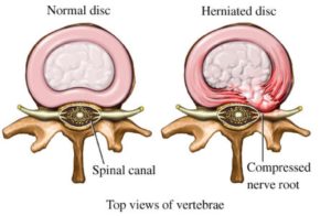
The Lumbar spine is composed for 5 boney building blocks that are called Vertebral Bodies. Sandwiched between each of the Vertebral Bodies is a Disc. The Disc is an important shock absorber that is composed of a thick outer layer and a gelatinous inner core. The Disc is susceptible to injury and degeneration. Common disc injuries include Disc Protrusions, Disc Herniations and Disc Extrusions.
The outer layer of the Disc known as the Annulus is rich is nerve fibers. Why does this matter? An increase in Disc pressure can stimulate these nerve fibers resulting in signficant pain and dysfunction. Lying down reduces the pressure on the Disc. Conversely sitting increases the pressure on the disc. Lower back pain when sitting can be due to Disc protrusions or Herniations.
Facet Injury
The Facet joint is a paired joint on the backside of the spine. They guide and limit motion of the bones in the low back (2). Like your knee and hip joints, the Facet joints are lined with cartilage which allows the joints to move smoothly. Facet joints are susceptible to injury and degeneration. Facet joint injuries can be caused by trauma, heavy lifting, repetitive movements and car accidents. Lumbar Facet joints are a common source of pain accounting for between 15-45% of low back pain (3).
Ligament Instability
Ligaments are thick pieces of connective tissue that connect bone to bone. They provide important stability for the low back. Important ligaments in the low back include the Supra and Interspinous Ligaments. Ligament laxity due to injury or cogential laxity can cause low back pain when sitting.
Nerve Compression
Nerves exit at each level in the low back. They exit through a boney doorway which is called the Neural Foramen. The doorway may be narrowed by Disc Protrusions, Disc Herniation, Facet overgrowth, Bone Spurs or Ligamental Thickening. This can cause significant irritation and or compression of the exiting nerve resulting in low back pain and Sciatica. Sitting increased the pressure on the Disc which in turn may increase the nerve compression. Often times the result is an increase in low back pain when sitting.
Spinal Stenosis
Stenosis refers to a narrowing. In the Lumbar spine there are two principal types of Stenosis: Central Canal Stenosis and Foraminal stenosis.
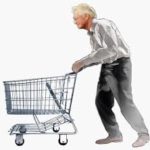
Central Canal Stenosis occurs when there is narrowing of the central spinal column causing pressure on the spinal cord. Patients with moderate to advanced Central Canal Spinal Stenosis tend to walk in hunched forward posture. This is often times referred to as a ” shopping cart sign” as patients are bent forward in an attempt to open up the narrowed canal by flexing forward. Patients can also have severe leg pain with walking causing them to stop after several yards due to pain.
Foraminal Stenosis occurs when there is narrowing of the boney doorway through which the spinal nerves exit. This can occur on one side or both sides. Symptoms can include radiating leg pain, numbness, weakness and muscle shrinkage.
Sacroiliac Joint Dysfunction
The Sacroiliac Joint (SIJ is a major joint in the low back. It is composed of the Sacrum and the part of your pelvis called the Ilium. There is an right and left SI joint. Like your knees and hips, the SI joint is lined with cartilage. It is stabilized through an extensive network of ligaments. The SIJ can be injured through both traumatic and nontraumatic causes (4). Traumatic causes of SI joint injury include sudden and repetitive heaving lifting, rear-end MVAs and hard falls onto the buttock. Non-traumatic causes include Lumbar Fusion, pregnancy due to elevated hormone levels and Scoliosis. SI joint injury and instability is common cause of unilateral low back pain when sitting.
Annular Tear
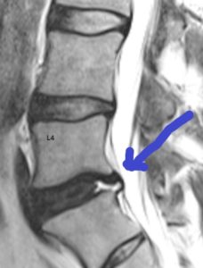
The Disc is composed of two principal parts: the Annulus Fibrous and the Nucleus Pulposus. The Nucleus is the gelatinous core that functions as a shock absorber. The Annulus Fibrous are outer fibers that surround and support the gelatious core. The Annulus is a strong radial tire-like structure that provides important stability for the Disc. The Annulus is susceptible in degeneration and injuries. A tear in the Annulus can occur with or without a Disc Protrusion or Herniation (5). The diagnosis of Annular tear is confirmed by MRI. On T2 imaging it is a bright small signal that looks a bright star in the evening sky. Annular tears are a common cause of low back pain when sitting.
Multifidus Muscle Atrophy
The stability of the Lumbar spine is dependent upon ligaments and muscles. The Multifidus is a small muscle that your doctor never talked to you. It is a critical to the stability of the Lumbar spine. Injury and weakness in the Multifidus can cause low back pain when sitting. To learn more please click on the video below.
Muscle Tightness
There a large number of muscles in the low back. They provide stability and allow us to rotate, bend forwards and backwards. They also have a protective role in that if there is an injury to the Lumbar Disc, Facet or tendon the muscles tighten. Persistent muscle tightness that does not respond to conservative treatment is a warning signal that you may have a low back injury. It warrants a deeper evaluation.
Spondylolisthesis
Spondylolisthesis is where one of the bones in the spine slips out of alignment. It can slip forward or backwards in relation to the other bones. This can cause injury to the Lumbar Discs, nerves and exiting nerves. Patients with Spondylolisthesis are unstable and can have lower back pain when sitting, bending or lifting.
Annular Tear
To understand annular tears, let us first review the anatomy of the spine. The lumbar spine is comprised of 5 boney building blocks called vertebral bodies. Sandwiched between the vertebral bodies are the lumbar discs. Each disc is comprised of an outer fibrous ring, the annulus fibrosis that surrounds the inner gelatinous center, which is called the nucleus. The disc absorbs the forces of daily living. The annulus has multiple layers of collagen that provide important support. The annulus is similar to the sidewall of a tire which provides important stability for the tire. Through trauma or degeneration, the outer annular fibers can become injured and or weakened.
Degenerative Scoliosis, also known as Adult-onset Scoliosis, is a medical condition that involves a side bending in the spine. The bending can be mild, moderate, or severe with side-bending to either the right or the left. The term degenerative means generalized wear and tear and is common as we get older. Degenerative scoliosis is the curvature of the spine that occurs as a result of degeneration of the discs, small joints, and building blocks. The Degenerative Scoliosis curve is often located in the low back and forms a ‘C” shape. There is a convex and a concave side. The convex side is the open side where it curves outward.
Disorders that affect and weaken the connective tissues such as tendons and ligaments. It is a hereditary disorder which means you are born with it. EDS has many different signs and symptoms which can vary significantly depending upon the type of EDS and its severity. It most commonly affects the skin, joints, and blood vessels. Joints are typically hypermobile with excessive joint range of motion because of a defect in collagen formation. In most cases Ehlers-Danlos syndrome is inherited. That is to say that you are born with it. The two main ways EDS is inherited are: autosomal dominant inheritance and autosomal recessive inheritance…
Injury or inflammation of the cervical facet can led to neck, shoulder and headache pain – called “cervical facet syndrome.” Cervical facet syndrome largely involves a joint in the posterior aspect of the cervical spine. It functions to provide stability and guide motion. cervical facet joint injection for cervical facet syndrome Cervical facet pain is common in patients who have sustained a whiplash injury, trauma to the neck or undergone cervical fusion. Physical examination is typically significant for restriction in range of motion along with pain. Each joint has a distinct referral pattern illustrated below. The Centeno-Schultz Clinic are experts at diagnosing and treating cervical facet dysfunction. Injury to the joint is not commonly detected by conventional radiographic studies.
A facet cyst, also known as a synovial cyst, is a fluid-filled sac that forms in the facet joint of the spine. The facet joints are small joints located between the vertebrae of the spine that provide stability and enable movement. Facet cysts typically develop due to degeneration and wear and tear of the facet joint, which can cause the joint capsule to stretch and weaken. This weakened capsule can then allow synovial fluid, which normally lubricates and nourishes the joint, to leak out and form a cyst. Facet cysts can cause various symptoms such as back pain, leg pain…
Failed Back Surgery Syndrome also called failed back is a clinical condition in which patients who have undergone low back surgery continue to have pain and dysfunction. Said another way the surgery that was intended to reduce pain and increase function FAILED. That is right, the surgery failed. You had the surgery, struggled with the pain postoperatively, diligently participated in physical therapy and yet the pain and limitation are still there. Unfortunately, this occurs frequently. Estimates range from 20-40% of patients who undergo low back surgery will develop Failed Back Surgery Syndrome. Pain is the most common symptom of Failed Back Surgery Syndrome…
Hamstring tendinopathy, a condition that causes pain and tenderness in the hamstring tendons, represents a common but often misunderstood musculoskeletal issue. It typically occurs in athletes and individuals engaged in high-intensity activities but isn’t limited to them. This condition, including its more specific form, high hamstring tendinopathy, can significantly impact daily and athletic performance, making understanding its nuances critical.
The sacroiliac joints reside between the sacrum (the tailbone segment of the spinal column) and the prominent wing-like iliac bones that form the pelvic girdle. There are two SI joints, one on the left and one on the right (highlighted in red in the image above), and along with the symphysis pubis joint at the front of the structure, they are critical for transferring forces and energy back and forth between the spine and the lower limbs. There are a number of reasons that an SI joint can become painful and inflamed, leading to SI joint syndrome. Trauma, such as a fall injury to the tail bone or a forced injury from a car accident for example, obviously can create problems in the SI joint…
Spinal instability is a condition that occurs when the spinal column is not able to maintain its normal alignment and function under normal loads. It can be caused by various factors such as trauma, degenerative changes, infections, tumors, or congenital abnormalities. In a stable spine, the bones, discs, ligaments, and muscles work together to support and protect the spinal cord and nerve roots. However, in an unstable spine, the structures that support the spine may be damaged or weakened. This can lead to abnormal movement and excessive stress on the spinal cord and nerves. In most cases, bone and joint problems…
Spinal stenosis is the narrowing of the central spinal canal and is a cause of significant pain and disability. Common causes of spinal stenosis include disc protrusion, facet overgrowth and ligamentum flavum thickening. Surgery is often chosen when conservative therapies fail despite the lack of convincing evidence that it is a superior treatment option. Are there alternatives to back surgery for spinal stenosis? Yes. Regenexx DDD utilizes precise platelet injections into the facets, muscles, and ligaments to treat the lumbar stenosis, treating all of the components of the issue, which is crucial. Spinal stenosis is often an age-related condition attributed…
Spondylolisthesis means that one vertebra is slipping forward or backwards on another. This causes the hole where the nerve exits (foramen) to get smaller (also called foraminal stenosis). It also causes more wear and tear on the facet joint which can lead to arthritis or what’s called “facet hypertrophy”.
spondylolisthesis recovery
The amount of slippage is graded 1-4, with grade 1 meaning that the one vertebra has slipped up to 25% on the other vertebra. Grade 2 means that one bone has slipped from 25-50% with higher grades indicating more slippage. The vast majority of patients are grade 1 to 2.
The thoracic spine, also known as the mid back, is that portion of the spine that is below the cervical spine (neck) and above the lumbar spine (low back). Thoracic spondylosis is a degenerative condition of the thoracic spine. The thoracic spine, also known as the mid back, is composed of many important different structures that work together to provide stability and movement. Thoracic spondylosis is a degenerative condition affecting the middle region of the spine, known as the thoracic spine. The major causes include: aging, genetics, poor posture, repetitive strain, and more. Symptoms can be mild, moderate, or severe…
The spinal discs are shock absorbers that live at each level between the vertebral bones (1). They have a tough outer annulus part and a soft inner gel part (nucleus pulposis). The outer covering can get damaged which can sometimes be seen on MRI and other times requires additional testing to identify. These tears are called: a torn disc, a disc tear, an annular tear, and when seen on MRI a “High-Intensity Zone” or HIZ. They can cause pain, mostly through ingrown nerves. There are torn disc findings that can be seen on MRI (HIZ) and these can be either asymptomatic (i.e. not painful) or…
Other Medical Conditions that Can Cause Lower Back Pain
Not all low back pain is due to injury of structures in the low back. Other important causes of low back pain include:
Kidney Stones
A Kidney Stone is a hard mass made from the chemicals in the urine. There are four types of Kidney Stones: Calcium Oxalate, Uric Acid, Struvite and Cystine. They can be small or large. They can localized irritation or block the flow or urine. Symptoms include severe one sided low back pain. Other symptoms include blood in the urine, fever and chills.
Gallbladder Disease
The Gallbladder is a part of your digestive system. Its main function is to store bile which helps break down fats. Inflammation of the Gallbladder or hardened deposits of bile called gallstones can cause Gallbladder disease and dysfunction. Symptoms include abdomen pain, fever, chills and low back pain. m
How You Can Prevent Lower Back Pain When You Sit (Overview)
The best way to avoid low back pain when sitting is to be proactive. Common strategies include:
Keep Moving
Movement is the best way to ward off back pain. Regular physical activity can make the back stronger to reduce future episodes of pain. Exercises should focus on increasing strength and improving range of motion.
Get Up from your Chair
Avoid prolonged sitting. If you sit at a desk in the office or at home all day, get up every 30 minutes and walk around. Consider a standing desk.
Posture
Optimize posture with neutral spinal alignment. Slouching compromises neutral spinal alignment and puts the Lumbar disc, facets, ligaments, and muscles at risk for injury and dysfunction.
Regular Exercise
Exercise has tremendous benefits both psychological and physical. Muscle mass and strength decline with age. Specifically, muscle mass decreases 3-8% per decade after the age of 30 and increases after the age of 60. Muscle strength is important for mobility and low back stability. Remember the adage: “Use it or lose it”
Stretching
Stretching has many important health benefits. Stretching can improve flexibility, range of motion, and posture which in turn can improve low back health and vitality.
Home Treatments to Ease Lower Back Pain
Mild low back pain after a long day or athletic event is not uncommon. When possible, it is best to resume normal or near-normal activity as soon as possible.
Conservative home treatment options include:
Intermittent Heat or Ice
Stretching
Gentle Massage
Safe oral anti-inflammatory medications such as fish oil and Turmeric. NSAIDs such as Motrin or Aleve have significant side effects and should be avoided.
Sleep with a pillow under your knees or on your side
When to Call Your Doctor
Pain tolerance can vary significantly from patient to patient. Low back pain that is tolerable for one patient may drive another one to the emergency room. You should consider seeing your doctor if you have:
Low back pain is persistent and not responsive to conservative care
Persistent leg numbness and tingling
Fever and chills
Loss of bowel or bladder function
How Important Is Getting A Solid Diagnosis?
Back pain is not a diagnosis. It is a symptom. For the very best clinical results, it is critical that an accurate diagnosis is made. Specifically based upon medical history, physical examination, and radiographic tests what structure or structures are causing your ongoing pain. This is the standard of care at the Centeno-Schultz Clinic.
Some pain clinics operate on the theory that a patient’s pain is arising from a single source. Many refer to this as identifying the “pain generator”. Accordingly, they will inject a specific part of the spine with diagnostic injections. For example, they may start with Lumbar Facet injections asking the question is the pain arising from the Facet Joints. If Facet Injections fail to provide benefit they move on and perform diagnostic Epidural injections. The problem with this model is that body is complex with many different, truly remarkable structures that all work in concert with one another.
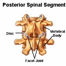
At the Centeno-Schultz Clinic we understand the importance of viewing the body as a whole. We view the spine as a functional spinal unit (FSU). The Functional Spine Unit is composed of Discs, Facet Joints, bones, ligaments, tendons, and muscles. To learn more about what all these parts do, please click on the video as it will bring this whole topic together.
Treatment Options
At the Centeno-Schultz Clinic we bring a standard of care and expertise that is truly unique. We were the first clinic in the world to inject Bone Marrow Concentrate into the Lumbar Disc in 2005. Treatment of common orthopedic conditions with PRP and Bone Marrow Concentrate is our singular focus. We do not inject Botox, burn nerves using Radiofrequency, use high dose steroids or manage chronic narcotics. We are committed to highest standard of care which includes:
Establishing the correct diagnosis
Using ultrasound or x-ray guidance on all injections
Treating the spine as a Functional Unit (FSU))
Determining the most appropriate treatment option: Platelet Rich Plasma or Bone Marrow Concentrate or both.
Determining the appropriate PRP dose given the injury and your age. To learn more please click here.
Determining the type of Bone Marrow Concentrate: same day procedure or culture expanded ( Regenexx- SD or Regenexx-C)*
Identifying the appropriate rehabilitation to ensure best clinical outcome.
The Centeno-Schultz Clinic are experts in the evaluation and treatment of low back pain when sitting. Our goal is to avoid patient’s reliance on potentially addicting narcotics or life altering surgeries. To learn more about the complications associated with low back fusions please click here.
In Summary
Low back pain is common and most likley with affect you at some point in your life.
The location of low back pain varies but is important to localize if possible.
The quality of low back pain also varies. Common examples include sharp, stabbing, dull, aching and burning.
Common causes of low back pain when sitting are disc protrusion, facet injury, ligament instability, nerve compression, spinal stenosis, annular tear and sacroiliac joint dysfunction.
Other important causes of low back that need to considered are kidney stones and gallbladder disease.
Strategies to prevention low back pain when sitting include limited sitting, improved posture, regular exercise and stretching.
It is time to contact a doctor when your low back is persistent despite conservative care, persistent numbness and tingling, fever and chills or loss of bowel or bladder function.
Low back pain is not a diagnosis but rather is a symptom. Getting an accurate diagnosis is critical if you want the very best clinical outcomes.
At the Centeno-Schultz Clinic treatment options for low back pain when sitting may include x-ray or ultrasound guided injections of PRP or bone marrow concentrate.
If you or a loved one has low back pain when sitting that has not been responsive to conservative care, please schedule a telephone Candidacy discussion with a board-certified, fellowship-trained physician. From the comfort of your home or office, learn what treatment options are available for you. Call today and stop the pain, misery, and suffering.
*Regenexx-C is only avialble at a licensed site in the Cayman Islands.
4.Chuang CW, Hung SK, Pan PT, Kao MC. Diagnosis and interventional pain management options for sacroiliac joint pain. Ci Ji Yi Xue Za Zhi. 2019 Sep 16;31(4):207-210. doi: 10.4103/tcmj.tcmj_54_19. PMID: 31867247; PMCID: PMC6905244.
5.Farshad-Amacker NA, Hughes AP, Aichmair A, Herzog RJ, Farshad M. Is an annular tear a predictor for accelerated disc degeneration? Eur Spine J. 2014 Sep;23(9):1825-9. doi: 10.1007/s00586-014-3260-8. Epub 2014 Mar 13. PMID: 24622958.
6.Volpi E, Nazemi R, Fujita S. Muscle tissue changes with aging. Curr Opin Clin Nutr Metab Care. 2004;7(4):405-410. doi:10.1097/01.mco.0000134362.76653.b2
FREE eBook Download (Click the Book Cover)
Ready to get help for your Lower Back Pain When Sitting?