Thoracic Compression Fracture
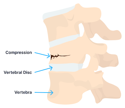
A compression fracture is a type of bone fracture that typically occurs in the vertebrae of the spine. It occurs when one or more of the vertebrae become compressed or crushed due to excessive forces or pressure.
The vertebrae are the bony building blocks that stack one upon another, forming the spine. Compression fractures most commonly occur in the vertebral body, which is the thick, rounded portion of each vertebra.
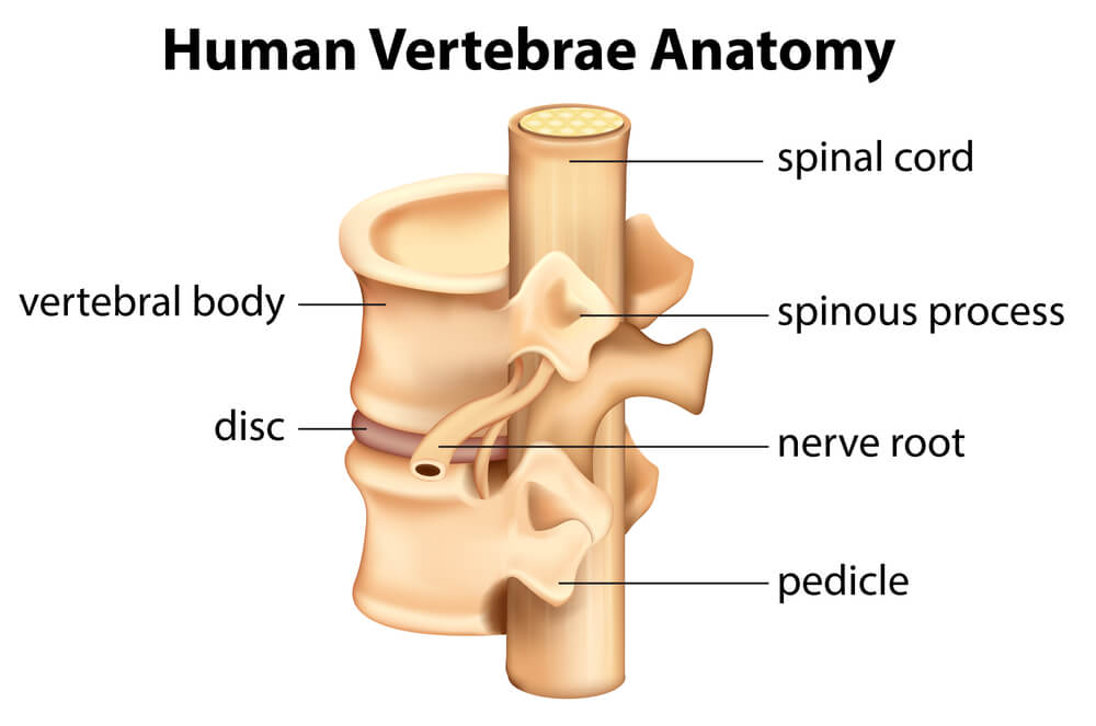
The spine is composed of three sections: cervical, thoracic, and lumbar. The cervical spine is also referred to as the neck, whereas the lumbar is the low back. The thoracic spine is that section of the spine below the neck and above the low back. It is commonly referred to as the mid back. A majority of the compression fractures occur in the thoracic spine.(1)
What Is a Thoracic Spine Compression Fracture?
A thoracic spine compression fracture is a break or crack in the thoracic vertebrae. It occurs most commonly in the vertebral body, which is the front portion of the vertebrae. Most occur in the lower thoracic spine, with 60-70% of thoracic fractures occurring in the T11-12 region.(2)
Thoracic compression fractures are common, with approximately 1.5 million per year in the United States. Approximately 25% of all postmenopausal women get a compression fracture during their lifetime.
What Causes the Condition?
As noted above, a thoracic compression fracture is a break or crack in the thoracic vertebrae, most often resulting in pain and dysfunction. The most common causes of thoracic compression fractures are:
Osteoporosis
This is the leading cause of compression fractures in the thoracic spine.(3) Osteoporosis is a medical condition characterized by the weakening of bones leading to reduced bone density and increased risk of fractures. Many factors contribute to the development of osteoporosis, including age, gender, hormonal changes, genetics, medication, alcohol, and lifestyle.
Trauma
A sudden, forceful impact to the back, such as from a fall, car accident, or sports injury can result in a compression fracture in the thoracic spine.
Age-Related
As we age, there is a reduction in bone density and spinal flexibility along with an increase in osteoarthritis and disc degeneration, which increase the risk of thoracic compression fractures.
Cancer Infiltration or Tumors
Tumors that originate in the spine or spread to the spine from other parts of the body (i.e. are metastatic) can weaken the vertebrae and increase the risk of compression fractures.
Osteopenia
This is a medical condition characterized by a reduction in bone mineral density, which weakens bones. The bone mineral density is lower than normal but not enough to be classified as osteoporosis. It represents a midpoint between normal bone density and osteoporosis. Risk factors for osteopenia include age, gender, nutritional factors, and lifestyle.(4)
Types of Compression Fractures
There are four major types of thoracic spine fractures. The two most common are:
- Compression fracture: This involves the collapse or compression of one or more vertebrae due to excessive force or pressure. It is a stable fracture and rarely accompanied by neurological injuries or deficits. In younger patients, it is typically the result of falls or motor vehicle collisions. In older patients, osteoporosis is the most common cause.
- Burst fracture: This is a severe type of vertebral fracture, characterized by the collapse or shattering of the vertebral body into multiple fragments. Fragments can irritate or compress the spinal cord or exiting nerve roots, leading to neurological injuries and deficits.
Thoracic Spine Compression Fracture Symptoms
Thoracic compression fractures can present with a variety of symptoms, ranging from mild to severe depending on the extent of the fracture and whether nearby structures are compromised. The most common symptoms include:
Chronic Back Pain
Pain is the most common symptom of thoracic compression fractures. The pain is typically localized to the middle or upper back. It can vary in intensity from mild to severe and typically is aggravated by coughing and sneezing.
Spinal Mobility Issues
Pain and stiffness associated with thoracic compression fractures can limit the range of motion in the spine and overall flexibility, making it difficult to bend or twist.
Development of a Hunched Posture
Thoracic compression fractures can lead to changes in spinal alignment and posture. Patients may develop excessive forward curvature (kyphosis) or a hunched posture.
Height Loss Due to Vertebral Compression
Thoracic compression fractures can cause a decrease in a person’s height due to the collapse or compression of the vertebral body. This loss of height may be noticeable over time and can contribute to changes in posture.
Nerve Complications
In some cases, thoracic compression fractures can irritate or compress the spinal cord or exiting nerve roots leading to numbness, tingling, and weakness in the limbs. In severe compression fractures, paralysis may occur if the spinal cord is injured.
Bladder and Bowel Control Challenges
Severe, untreated fractures can manifest in problems controlling bladder or bowel functions. Severe compression fractures that affect the lower thoracic spine may compress the spinal cord and disrupt nerve signals responsible for bowel and bladder function, leading to incontinence or difficulty with urination or defecation.
How Is This Condition Diagnosed?
The diagnosis of a thoracic compression fracture involves a combination of medical history, physical examination, and imaging studies. The most common imaging studies include:
- Spine X-ray: This is often the first imaging study used to evaluate thoracic compression fractures. X-rays can show changes in the shape of the vertebral body, loss of height, and other signs suggestive of a thoracic compression fracture.
- CT scan: A CT scan provides detailed cross-sectional images of the chest and can accurately detect and evaluate fractures.
- MRI: MRI provides detailed images of bone and soft tissue structures such as the spinal cord, nerves, muscles, tendons, and ligaments.
- Dual-energy X-ray absorptiometry (DEXA): DEXA is the gold standard for the diagnosis of thoracic compression fractures because it can measure central bone mass and has excellent specificity.
- Bone density testing: Bone density testing evaluates the density and strength of the bones in the thoracic region and helps assess the severity of such fractures.
- Three-phase bone scan: This is a valuable diagnostic tool for identifying thoracic compression fractures and their severity. Its three-step process provides detailed information on blood flow, skeletal integrity, and the overall bone health of the thoracic region.
Conservative Treatment Options
Conservative treatment options for thoracic compression fractures aim to manage pain, promote healing, and prevent further complications without surgical intervention. These approaches may include:
- Bed rest/activity limitation: This is essential, as it allows for the healing of fractures.
- Bracing: Bracing helps to stabilize the spine, reduce pain, and provide proper alignment during the healing process.
- Pain management: In addition to medication, techniques such as heat, ice, massage, and TENS units may provide relief from pain and muscle tightness.
The Centeno-Schultz Approach to Treating Thoracic Spine Compression Fractures
At the Centeno-Schultz Clinic, we take a global approach to the spine, acknowledging that the many different moving parts must work in a coordinated and dynamic fashion.
In the case of thoracic compression fracture, other spinal structures are affected and may be responsible for pain, muscle spasms, and dysfunction. Examples include the spinal ligaments, discs, facet joints, tendons, and ligaments. Each of these structures warrants evaluation and possible treatment in cases involving thoracic compression fractures.
To learn more about our unique approach to the spine, please click here:
Physical Therapy
Physical therapy can play a crucial role in managing thoracic compression fractures. It can aid in pain relief, improving mobility, and restoring strength and function through targeted exercises, manual therapy, and other specialized interventions.
Prolotherapy Injections
It has been successful in the treatment of many disorders including neck, shoulder, knee, and ankle pain. Dr. Centeno recently published an article in The Journal of Prolotherapy in which he discusses the use of x-ray guidance with prolotherapy. This ensures that the injection is in the correct place to maximize clinical results. Dr. Centeno discusses the use of prolotherapy for the treatment of neck, knee, sacroiliac joint, ankle, ischial tuberosity, and shoulder pain. At the Centeno-Schultz Clinic x-ray guided prolotherapy is just one of the therapies utilized in the successful treatment of pain. Regenerative injection therapy (RIT) or prolotherapy…
Read More About Prolotherapy InjectionsPRP Injections
PRP is short for platelet-rich plasma, and it is autologous blood with concentrations of platelets above baseline values. The potential benefit of platelet-rich plasma has received considerable interest due to the appeal of a simple, safe, and minimally invasive method of applying growth factors. PRP treatments are a form of regenerative medicine that utilizes the blood healing factors to help the body repair itself by means of injecting PRP into the damaged tissue. In regenerative orthopedics, it is typically used for the treatment of muscle strains, tears, ligament and tendon tears, minor arthritis, and joint instability. There have been more than 30 randomized controlled trials of PRP…
Read More About PRP InjectionsPreventive Measures for Optimum Spine Health
A thoracic compression fracture can be a debilitating injury that compromises function and quality of life. Preventive measures are important and focus on promoting bone health, reducing the risk of falls, and minimizing factors that contribute to bone loss.
Maintain Bone Health:
- Ensure adequate calcium and vitamin D intake.
- Get regular exercise: engage in weight-bearing and muscle-strengthening exercises regularly to promote bone density and strength.
- Avoid smoking.
- Avoid excessive alcohol consumption.
Fall Prevention:
- Maintain a safe environment.
- Use assistive devices such as canes or walkers, if needed.
- Improve balance and strength.
- Get regular exercise.
Osteoporosis Screening and Management:
- Ask your medical practitioner for a bone density test.
- Discuss osteoporosis treatment with your medical practitioner.
- Ensure you:
- Fuel up with bone-loving nutrients.
- Know your bone density.
- Medicate wisely.
- Kick the smoking habit.
- Drink responsibly.
What Happens When a Thoracic Spine Compression Fracture Is Left Untreated?
A thoracic compression fracture is a serious medical condition that, if left untreated, can potentially cause significant health issues. The most common complications and long-term consequences include:
- Chronic pain: A fracture is but one of the possible causes of pain. Other potential sites include the disc, facet joints, ligaments, and tendons.
- Reduced mobility: An untreated thoracic spine compression generally leads to reduced mobility.
- Change in posture: Compression fractures can cause the affected vertebrae to collapse, leading to a forward curvature of the spine known as kyphosis.
- Neurological complications: Complications include lower extremity weakness, and bowel and bladder dysfunction as a result of compression of the spinal cord or exiting nerve roots.
- Decrease in quality of life: A decrease in quality of life can occur, due to pain, limited mobility, and possible neurologic symptoms.
Treat Thoracic Spine Compression Fracture with the Centeno-Schultz Clinic
Thoracic compression fractures occur most frequently in the lower thoracic region, most commonly occurring in the T11-12 level. Thoracic compression fractures are common, with approximately 1.5 million per year in the United States.
The most common causes are osteoporosis, trauma, osteopenia, and tumors. Symptoms vary depending on the severity of the fracture and include pain, reduced mobility, compromised posture, loss of height, and potential nerve dysfunction resulting in bladder and bowel dysfunction.
A comprehensive approach to the spine and compression fracture is essential for the best clinical outcomes and results. The spine has many moving parts that work together in a synchronized fashion, and each of these should be evaluated.
If left untreated, a thoracic compression fracture can lead to chronic pain, limited mobility, changes in posture, potential neurologic complications, and a compromise in quality of life.
Are you a Candidate?

John Schultz, MD
John R. Schultz M.D. is a national expert and specialist in Interventional Orthopedics and the clinical use of bone marrow concentrate and PRP for orthopedic injuries. He is board certified in Anesthesiology and Pain Medicine and underwent fellowship training. Dr. Schultz has extensive experience with same day as well as culture expanded bone marrow concentrate and sees patients at the CSC Broomfield, Colorado Clinic, as well the Regenexx Clinic in Grand Cayman. Dr. Schultz emphasis is on the evaluation and treatment of thoracic and cervical disc, facet, nerve, and ligament injuries including the non-surgical treatment of Craniocervical instability (CCI).
Other Resources for Thoracic Compression Fracture
-
The C1 And C2 Vertebrae – What To Know
The C1 and C2 vertebrae, also known as the atlas and axis, are the uppermost bones in the spinal column. They play a crucial role in supporting the skull and enabling head movements. Damage or injury to these vertebrae often leads to pain, limitations to daily activities, and reduced quality of life. Many patients, without…
-
Spinal Fusion Recovery: What to Expect
Navigating spinal fusion recovery can be a daunting prospect, given its impact on daily life and mobility. Understanding what to expect during this process is crucial for individuals undergoing this procedure. In this article, we’ll explore the typical timeline, challenges, and strategies for managing recovery after spinal fusion surgery, providing insights to help individuals prepare…
-
Back Fusion
Spinal fusion, also known as back fusion, is a surgical procedure designed to help severe spinal instability that causes severe pain or nerve injuries. It involves permanently connecting two or more vertebrae in your spine to eliminate motion between them. This article will delve into the intricacies of spinal fusion, exploring the reasons behind the…
-
Understanding the Thoracic and Lumbar Spines
The thoracic spine and lumbar spine make up a vital nexus of stability and mobility in the human body. In this exploration, we delve into the biomechanics and complexities that define these regions, unraveling their significance in posture, movement, and overall well-being. Understanding the thoracic and lumbar spine not only illustrates the mechanics of our…
-
The L5 Vertebra: Everything You Need to Know
The spine, consisting of 33 individual bones called vertebrae, is divided into five major regions: cervical (neck), thoracic (upper back), lumbar (lower back), sacral (pelvic region), and coccygeal (tailbone). These vertebrae are separated by intervertebral discs, which act as cushions and allow for flexibility in movement. The lumbar spine, also known as the lower back,…
-
Exploring the Lumbosacral Joint: What Should You Be Wary Of?
The L5 S1 joint, or the lumbosacral joint, is a critical joint. It lies between the lumbar segment of the spine and the auricular processes of the sacrum. The L5-S1 joint plays a vital role in transmitting the weight of the body via the sacrum and ilium downwards. The weight is distributed to the femur…
-
Symptoms of Thoracic Herniated Disc
Your mid back pain has been unrelenting since the accident. Rest, medications, and physical therapy have failed to provide significant or sustained benefits. Your doctor thinks you have a thoracic disc herniation and thinks you have a thoracic herniated disc. What are the symptoms? And what can you do? Let’s dig in. What Is A…
-
Thoracic Spine MRI: What is It?
Are you getting an MRI of your thoracic spine? Get the help you need with the Centeno-Schultz Clinic. Learn what you need to know about this exam. What to expect on your thoracic spine MRI scan? What exactly is an MRI of the thoracic spine? How does an MRI work? What are the risks and…
-
Could The Anterior Longitudinal Ligament Be Causing Your Neck Pain?
The neck pain is unrelenting, and it all started after being rear-ended at a stoplight. Spine X-rays demonstrated some abnormal movement of the spinal bones. Both conservative care and medications have failed. Your doctor thinks you have injured a spinal ligament. What is the anterior longitudinal ligament? What is its role? What ligaments are injured…
References
- Alexandru D, So W. Evaluation and management of vertebral compression fractures. Perm J. 2012 Fall;16(4):46-51. doi: 10.7812/TPP/12-037. PMID: 23251117; PMCID: PMC3523935.
- Wong CC, McGirt MJ. Vertebral compression fractures: a review of current management and multimodal therapy. J Multidiscip Healthc. 2013 Jun 17;6:205-14. doi: 10.2147/JMDH.S31659. PMID: 23818797; PMCID: PMC3693826.
- Madassery S. Vertebral Compression Fractures: Evaluation and Management. Semin Intervent Radiol. 2020 Jun;37(2):214-219. doi: 10.1055/s-0040-1709208. Epub 2020 May 14. PMID: 32419735; PMCID: PMC7224975.
- Pouresmaeili F, Kamalidehghan B, Kamarehei M, Goh YM. A comprehensive overview on osteoporosis and its risk factors. Ther Clin Risk Manag. 2018 Nov 6;14:2029-2049. doi: 10.2147/TCRM.S138000. PMID: 30464484; PMCID: PMC6225907.