
Neck pain is a significant contributor to worldwide disability, affecting people of all ages and both sexes. According to the Global Burden of Disease Study 2019, neck pain is the fourth leading cause of years lived with disability (YLD) globally, accounting for 43.9 million YLDs.
Neck pain can be caused by various factors such as poor posture, trauma, degenerative changes, and inflammation. It can also lead to other health problems such as headaches, shoulder pain, and upper back pain, which can further affect an individual’s quality of life.
In medical terms, neck pain is described as pain originating in the cervical spine. So today, let’s dive into the details of the cervical spine so you can have a better understanding of what is causing your neck pain!
Spinal Anatomy: Bones, Joints, And Ligaments
The cervical spine is a construction of bones (vertebrae), joints (facet joints), cushions (intervertebral discs), ligaments (connect bone to bone), and muscles.
All these structures are put together in an intelligent design to allow all the ranges of motion of your neck while protecting the spinal cord in the center of the spine.
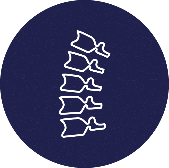
The Vertebrae

There are 7 cervical vertebrae and each has its own design to allow motion while protecting your nervous tissue.
Atlas And Axis
The atlas and axis are the first two vertebrae in the cervical spine, located at the top of the neck. They have unique features that enable them to perform specific functions.
The atlas (C1) is the topmost vertebra in the spine, and its primary function is to support the weight of the head. The atlas lacks a vertebral body and has a ring-like structure that allows for the passage of the spinal cord. It also has two large bony protrusions called lateral masses that articulate with the occipital condyles of the skull, allowing for the nodding movement of the head.
The axis (C2) is the second vertebra in the spine and has a unique structure that allows for rotation of the head. It has a bony protrusion called the odontoid process or dens that extends upward from the vertebral body of the axis and articulates with the atlas. The dens acts as a pivot point, allowing the head to rotate from side to side.
Together, the atlas and axis play an essential role in supporting the weight of the head, allowing for nodding and rotation movements, and protecting the spinal cord. They also provide stability to the upper cervical spine and facilitate the movement of the neck.
C3-C7 Vertebrae
The C3 to C7 vertebrae are the five vertebrae that make up the lower part of the cervical spine, located in the neck region. Each of these vertebrae have unique features that enable them to perform specific functions.
The C3 vertebra provides support and stability to the cervical spine, connecting the upper and lower cervical vertebrae.
The C4 vertebra plays a role in neck extension, allowing the head to tilt backward.
The C5 vertebra is responsible for neck flexion, enabling the head to bend forward.
The C6 vertebra facilitates the bending of the neck to the side, allowing the head to tilt laterally.
The C7 vertebra is the largest and strongest of the cervical vertebrae, providing support and stability to the neck. It also has a bony prominence at its base called the vertebra prominens, which is visible and palpable on the back of the neck.
Collectively, the C3 to C7 vertebrae play a crucial role in supporting the weight of the head, facilitating neck movement, protecting the spinal cord, and allowing for the passage of nerve roots that control movement and sensation in the upper body. Any damage or dysfunction in these vertebrae can lead to neck pain, stiffness, and other related symptoms.
Intervertebral Discs

Intervertebral discs are fibrocartilaginous cushions located between adjacent vertebrae in the spine.
They have several important functions, including:
- Shock absorption: The intervertebral discs act as shock absorbers, helping to distribute and dissipate the forces that occur during spinal movements such as walking, running, and jumping.
- Load-bearing: The discs help to support the weight of the upper body and maintain the spacing between the vertebrae, which allows for proper spinal alignment.
- Movement facilitation: The intervertebral discs allow for the movement of the spine, including flexion, extension, lateral bending, and twisting.
- Nerve protection: The discs help to protect the spinal nerves that exit the spinal cord and travel to various parts of the body.
- Nutrition supply: The intervertebral discs do not have their own blood supply, but they receive nutrients and oxygen through a process called diffusion, which occurs when the discs undergo compression and relaxation during spinal movements.
Overall, the intervertebral discs play a crucial role in maintaining the health and function of the spine, and any damage or degeneration to these discs can lead to various spinal conditions such as herniated discs, degenerative disc disease, and spinal stenosis.
The intervertebral discs are made up of a complex structure of different tissues that work together to provide the disc’s unique properties.
- The disc’s central portion, called the nucleus pulposus, is a gel-like substance that provides cushioning and shock-absorbing properties.
- The outer layer, called the annulus fibrosus, is made up of tough fibrous tissue that surrounds and encases the nucleus pulposus.
The nucleus pulposus is composed of water, collagen fibers, and proteoglycans. Proteoglycans are large molecules that attract water, giving the nucleus pulposus its gel-like consistency. Collagen fibers provide strength and stability to the nucleus pulposus.
The annulus fibrosus is made up of several layers of collagen fibers, arranged in a criss-cross pattern. The fibers are strongest near the center of the disc and gradually weaken towards the outer layers. This structure helps to distribute and absorb the forces placed on the disc during spinal movement.
The intervertebral disc also contains blood vessels and nerves that supply the disc’s outer layers, but the nucleus pulposus does not have its own blood supply. The disc receives its nutrition through a process called diffusion, which occurs when the disc undergoes compression and relaxation during spinal movement.
Overall, the complex structure of the intervertebral disc provides it with unique properties that allow it to function as a shock absorber, load-bearing structure, and facilitator of spinal movement.
Supporting Structures Of The Spine
Above are the foundation of the spine and then layers start to develop that allow the function of the spine.
Muscles
Movement of the spine is made possible by all the muscles that attach to the spine!

Anterior (front) Neck Muscles
The anterior cervical muscles are a group of muscles located in the front of the neck. These muscles play an essential role in supporting and stabilizing the head and neck and facilitating neck movements. The major anterior cervical muscles include:
- Sternocleidomastoid (SCM): The SCM is a large muscle that runs from the base of the skull to the sternum and clavicle. It allows for rotation of the head and neck to the opposite side, flexion of the neck, and extension of the head.
- Scalene muscles: The scalene muscles are a group of three muscles (anterior, middle, and posterior) that run from the cervical vertebrae to the first two ribs. They play a role in elevating the first and second ribs during inhalation and rotation of the neck.
- Longus colli muscle: The longus colli muscle is a deep muscle that runs along the front of the vertebral column from the skull to the thorax. It is responsible for flexing the neck and stabilizing the cervical spine.
- Longus capitis muscle: The longus capitis muscle is a deep muscle that runs from the skull to the cervical vertebrae. It is responsible for flexing the head and neck and stabilizing the cervical spine.
Overall, the anterior cervical muscles play a crucial role in maintaining proper posture, stabilizing the head and neck, and facilitating neck movements. Dysfunction or weakness in these muscles can lead to neck pain, stiffness, and other related symptoms.
Prevertebral Neck Muscles
The prevertebral neck muscles are a group of muscles located in the front of the spine, just deep to the cervical spine. They are responsible for stabilizing the cervical spine, supporting the head and neck, and facilitating neck movements. The major prevertebral neck muscles include:
- Longus colli muscle: The longus colli muscle is a deep muscle that runs along the front of the vertebral column from the skull to the thorax. It is responsible for flexing the neck and stabilizing the cervical spine.
- Longus capitis muscle: The longus capitis muscle is a deep muscle that runs from the skull to the cervical vertebrae. It is responsible for flexing the head and neck and stabilizing the cervical spine.
- Rectus capitis anterior muscle: The rectus capitis anterior muscle is a small muscle that runs from the base of the skull to the atlas (C1) vertebra. It is responsible for flexing the head and neck.
- Rectus capitis lateralis muscle: The rectus capitis lateralis muscle is a small muscle that runs from the transverse process of the atlas (C1) vertebra to the base of the skull. It is responsible for tilting the head to the side.
- Anterior scalene muscle: The anterior scalene muscle is one of the three scalene muscles located in the front of the neck. It runs from the cervical vertebrae to the first rib and is responsible for elevating the first rib and assisting with neck flexion.
Overall, the prevertebral neck muscles work together with the anterior cervical muscles to maintain proper posture, support the head and neck, and facilitate neck movements. Dysfunction or weakness in these muscles can lead to neck pain, stiffness, and other related symptoms.
Ligaments
The spine has several ligaments that provide support and stability to the vertebral column. The major ligaments of the spine include:
- Anterior longitudinal ligament (ALL): The ALL runs along the anterior aspect of the vertebral bodies, from the skull to the sacrum. It helps to prevent excessive extension of the spine and limits lateral flexion and rotation.
- Posterior longitudinal ligament (PLL): The PLL runs along the posterior aspect of the vertebral bodies, within the vertebral canal. It helps to prevent excessive flexion of the spine and limits lateral flexion and rotation.
- Ligamentum flavum: The ligamentum flavum is a strong, elastic ligament that connects the laminae of adjacent vertebrae. It helps to maintain the stability of the spinal column and assists with spinal movement.
- Interspinous ligament: The interspinous ligament connects the spinous processes of adjacent vertebrae. It helps to limit flexion and rotation of the spine.
- Supraspinous ligament: The supraspinous ligament runs along the tips of the spinous processes, from the cervical to the sacral region. It helps to limit flexion and assists with spinal extension.
- Intertransverse ligament: The intertransverse ligament connects the transverse processes of adjacent vertebrae. It helps to limit lateral flexion of the spine.
Overall, the ligaments of the spine work together to provide stability and support to the vertebral column, preventing excessive movement and maintaining proper alignment. Dysfunction or damage to these ligaments can lead to instability, spinal deformities, and related symptoms.
Joints
The spine has several types of joints that allow for movement and provide support and stability to the vertebral column. The major joints of the spine include:
- Intervertebral joints: The intervertebral joints are the joints between adjacent vertebral bodies, separated by intervertebral discs. These joints allow for flexion, extension, lateral bending, and rotation of the spine.
- Facet joints: The facet joints are the joints between the superior and inferior articular processes of adjacent vertebrae. These joints allow for gliding and sliding movements of the spine and play a role in stabilizing the vertebral column.
- Atlanto-occipital joint: The atlanto-occipital joint is the joint between the atlas (C1) vertebra and the occipital bone of the skull. This joint allows for nodding movements of the head.
- Atlantoaxial joint: The atlantoaxial joint is the joint between the atlas (C1) and axis (C2) vertebrae. This joint allows for rotation of the head and neck.
- Costovertebral joints: The costovertebral joints are the joints between the ribs and the thoracic vertebrae. These joints allow for expansion and contraction of the rib cage during breathing.
Overall, the joints of the spine work together to provide flexibility, stability, and support to the vertebral column, allowing for a range of spinal movements and protecting the spinal cord. Dysfunction or damage to these joints can lead to pain, stiffness, and related symptoms.
Its Role In The Central Nervous System
All the above structure are designed for 2 main things
- Protect the CNS = Central Nervous System = Spinal cord and its nerve roots
- Allow mobility and movement (while protecting the CNS)
Common Spinal Problems In The Cervical Region
In the United States, neck and back pain are also major causes of disability, affecting millions of individuals each year. According to the National Institute of Neurological Disorders and Stroke, back pain is the second leading cause of disability among adults in the United States, and neck pain is also a significant contributor to disability.
The impact of neck and back pain on disability can be significant, affecting individuals’ ability to work, perform daily activities, and engage in leisure activities. It can also lead to related health problems such as depression, anxiety, and reduced quality of life.
Imaging-Get movement based imaging (DMX or Upright MRI with flexion and extension).

Get Typed via a Telemed Appointment-There are 8 different types of CCI based on which ligaments are Injured.
Exam-If you’re a candidate for precise orthobiologic injections, you fly in for an exam and treatment. A hands-on exam refines what we need to treat.
Treatment-Get treated with a CCI focused, orthobiologic treatment plan tailored to you. That includes PICL or whichever image guided, precise injections are the most likely to help you.
Diagnosing Spinal Conditions
With so many parts and each able to create certain symptoms and signs on examination. It is critical if you are experiencing neck or back pain that you have a good examination to identify what is what…. And NO the MRI will not tell you where your pain is coming from.
While MRI images help physicians put the puzzle together with your specific symptoms and examination, it will NOT tell you where the pain is coming from! For that, you need a doctor to listen to your detailed history, perform a detailed examination and then refer to imaging to confirm.
If a doctor takes 10 minutes to evaluate you, or never examines you and then only reviews your MRI, then you are at the wrong physician, and it is unlikely they will help you get better!
Common Treatments For Spinal Conditions In The Cervical Area
There are always some basics when it comes to your spine health:
- Medications / supplementation
- Lifestyle Adjustments
- Physical Therapy
With the goals to avoid:
- Corticosteroid injections / Radiofrequency Ablations
- Spinal Surgeries
- Fusions
- ADR = artificial disc replacements
- Laminectomies
- Discectomies
Regenexx® Alternatives to Back & Neck Surgery
If simple and basic things as mentioned above aren’t helping your pain, then taking a holistic approach with ortho-biologics may be the answer! We utilize the most advanced biologics on the affected areas through a Functional Spinal Unit (FSU) approach.
Watch the following presentation to understand more:
How To Protect Your Spine
In addition to biologics / therapy and regular activity here are some other good tips for maintaining good overall spine health:
- Maintain good posture
- Take frequent breaks
- Use ergonomic equipment
- Avoid carrying heavy l oads on your head or shoulders
- Exercise regularly
- Avoid activities that strain the neck
The Vital Importance Of Spine Health
If you’re someone that is active and plans on continuing that lifestyle well into your later years, then maintaining spinal health is paramount to achieving that! If you have been dealing with spine issues, contact our office to set up a full evaluation where one of our experts will do an in-depth evaluation and review your best options to keep you moving forward!
Consult with us whether you are experiencing cervical spine pain or just want to know about your overall spinal health. We can help.