
The neck is a vital and complex part of the human anatomy, supporting the head and facilitating essential functions like movement, breathing, and communication. When neck pain, injuries, or other symptoms arise, healthcare providers often turn to diagnostic tools such as neck X-rays to assess the condition of the cervical spine and surrounding structures.
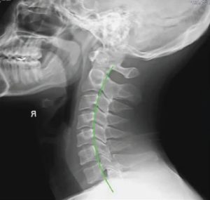
Healthy neck vs unhealthy neck
In this article, we will delve into the world of neck X-rays, exploring their purpose, preparation, procedure, and the significance of the results.
Whether you’re curious about what to expect during a neck X-ray or you’ve been advised to undergo one, this comprehensive guide will provide you with the information you need to understand this diagnostic procedure and its implications for your neck health.
Learning About the Neck
The neck is a vital and complex part of the human anatomy that connects the head to the rest of the body. It plays a crucial role in supporting the head, facilitating movement, and housing essential structures such as the spinal cord, blood vessels, and airways.
Let’s delve into its anatomy, structure, the risks of damage, and the effects of different factors on the neck:
Anatomy of the Neck
- Vertebrae: The neck consists of seven cervical vertebrae, labeled C1 to C7, which form the cervical spine. These vertebrae provide structural support and protect the spinal cord.
- Muscles: The neck is surrounded by numerous muscles that enable various movements of the head, such as turning, tilting, and nodding. Key muscles include the sternocleidomastoid, trapezius, and scalene muscles.
- Blood vessels: Major blood vessels, including the carotid arteries and the jugular veins, pass through the neck. The carotid arteries supply blood to the brain, while the jugular veins drain blood from the head.
- Larynx and trachea: The neck houses the larynx (voice box) and trachea (windpipe), which are essential for breathing and vocalization.
- Esophagus: The esophagus, responsible for transporting food from the mouth to the stomach, also passes through the neck.
Structure of the Neck
The neck comprises a complex arrangement of bones, muscles, ligaments, tendons, blood vessels, nerves, and other tissues. The cervical vertebrae provide stability and protection for the spinal cord, while the surrounding muscles and soft tissues enable mobility and support various functions.
Risks of Damage to the Neck
- Trauma: The neck is vulnerable to various forms of trauma, such as whiplash from car accidents, sports injuries, or falls. Trauma to the neck can lead to fractures, dislocations, or soft tissue injuries.
- Neck pain and strain: Poor posture, excessive use of electronic devices, or heavy lifting can lead to neck pain and muscle strain.
- Neck disorders: Conditions like cervical spondylosis, herniated discs, and cervical stenosis can affect the neck’s structure and function, leading to pain and reduced mobility.
- Infections: Infections in the neck, such as abscesses or tonsillitis, can cause localized swelling, pain, and discomfort.
Effects of Different Factors on the Neck:
Several factors can affect the neck and its health:
- Poor posture: Prolonged poor posture, like hunching over a computer, can strain neck muscles, leading to discomfort and potential long-term problems.
- Aging: As people age, the cervical spine may undergo degenerative changes, such as the development of osteoarthritis, which can result in neck pain and reduced mobility.
- Obesity: Excess body weight can place additional strain on the neck’s supporting structures and exacerbate neck pain.
- Injury: Neck injuries can vary in severity, ranging from minor muscle strains to severe spinal cord damage. The effects of an injury depend on the type and location of the damage.
- Lifestyle choices: Smoking and poor nutrition can contribute to the development of conditions that affect the neck, such as atherosclerosis (narrowing of arteries) and osteoporosis (weakening of bones).
The neck is a critical anatomical region with a complex structure that supports various functions, including head movement, breathing, and speech. It is susceptible to a range of risks and can be affected by various factors, leading to pain, injury, or functional limitations. Correct posture, exercise, and preventive measures can help maintain neck health and reduce the risk of damage.
What Is an X-Ray of the Neck?
An X-ray of the neck, often called a cervical spine X-ray, is a medical imaging procedure used to visualize the bones and some soft tissues in the neck region. This diagnostic tool allows healthcare professionals to assess the structure and alignment of the cervical spine, which consists of the seven cervical vertebrae (C1 to C7) and their associated structures.
Why Would Anyone Need an X-Ray?
The purpose of a neck X-ray includes some of the following:
- Diagnosis: A neck X-ray is commonly used to diagnose various conditions and injuries that affect the cervical spine. These include fractures, dislocations, degenerative changes, and abnormalities of the vertebrae.
- Trauma assessment: After a neck injury, such as a car accident or fall, a neck X-ray may be performed to evaluate for any bone fractures or other traumatic injuries that may require immediate medical attention.
- Arthritis and degeneration: X-rays of the neck can help diagnose conditions like cervical spondylosis, which is characterized by degenerative changes in the cervical spine, including the development of bone spurs and narrowing of the spinal canal.
- Alignment issues: Doctors use neck X-rays to assess the alignment of the cervical vertebrae and to look for conditions like scoliosis or abnormal curvature of the spine.
Walking Through the Procedure
Obtaining a neck X-ray is a common medical procedure used to diagnose various conditions related to the cervical spine and neck region. Understanding what to expect before, during, and after the procedure can help alleviate any concerns. Here, we’ll walk you through the process step by step.
Preparing for a Neck X-Ray
Before undergoing a neck X-ray, there are several essential preparations to keep in mind:
- Medical history: You may be asked about your medical history, including any previous neck injuries, surgeries, or existing conditions. Inform your healthcare provider if you are pregnant or suspect you might be, as alternatives to X-rays may be considered.
- Clothing and jewelry: You’ll typically be provided with a hospital gown to wear during the procedure. Remove any clothing or jewelry that could interfere with the X-ray images, especially around the neck area.
- Radiation safety: Understand that X-rays involve exposure to ionizing radiation, which carries a small risk. Your healthcare provider will take precautions to minimize radiation exposure, such as using a lead apron and collar to shield other parts of your body.
What to Expect
During a neck X-ray, here’s what you can expect:
- Positioning: You will be positioned by a radiologic technologist in front of the X-ray machine. Depending on the specific X-ray views required, you may be asked to stand, sit, or lie on an X-ray table.
- Immobilization: To ensure clear and accurate images, you’ll be asked to remain as still as possible during the X-ray exposure. In some cases, you may need to hold your breath briefly.
- Multiple views: Your healthcare provider may take X-rays from different angles and positions to get a comprehensive view of the cervical spine. This may involve turning your head or neck into different positions.
- Duration: X-ray exposures are typically brief, lasting only a few seconds to a minute, depending on the complexity of the images required.
During the Procedure
Here are some specific details about what happens during the neck X-ray procedure:
- X-ray machine: The radiologic technologist will position the X-ray machine as needed to target the cervical spine area.
- Lead shielding: You may be provided with lead shielding, such as a lead apron or collar, to protect other body parts from unnecessary radiation exposure.
- Communication: You’ll be in communication with the technologist throughout the procedure. They may instruct you on when to hold still, breathe, or change positions.
- Minimal discomfort: The X-ray itself is painless, and you should not feel any discomfort from the radiation exposure.
- Post-procedure: Once the necessary images have been obtained, you will be allowed to dress and return to your regular activities unless further medical evaluation is needed based on the X-ray findings.
Remember that the specific details of your neck X-ray procedure may vary, depending on the healthcare facility, the equipment used, and the purpose of the X-ray. It’s essential to follow any instructions from your healthcare provider or the radiologic technologist to ensure a successful and safe imaging procedure.
Reading the Results of Your Neck X-Ray
Once you’ve undergone a neck X-ray, the next step is interpreting the results. These results can provide valuable insights into the health and condition of your cervical spine. Let’s explore the two possible outcomes: when your neck X-ray results are normal and when they are abnormal.
When Your Neck X-ray Results Are Normal
If your neck X-ray results are normal, it means that no significant abnormalities or issues were detected during the imaging. This is typically good news and suggests that the structures of your cervical spine, such as the vertebrae, discs, and joints, appear to be within the normal range. However, it’s essential to remember that a normal X-ray does not necessarily rule out all potential neck-related problems or conditions, especially those involving soft tissues or nerve issues.
When Your Results Are Abnormal
If your neck X-ray results are abnormal, it indicates that there may be issues or abnormalities in your cervical spine. Abnormal findings on a neck X-ray can include:
- Fractures: A fracture or break in one or more cervical vertebrae may be evident on the X-ray images. The severity and location of the fracture will determine the appropriate treatment.
- Dislocations: An X-ray can reveal if any cervical vertebrae are out of their normal alignment, indicating a dislocation. This may require immediate medical attention to realign the bones.
- Degenerative changes: X-rays can show signs of degeneration, such as the development of bone spurs or narrowing of the spinal canal. These findings suggest conditions like cervical spondylosis.
- Tumors or abnormal growths: In some cases, X-rays may reveal masses or abnormal growths within the cervical spine, which may require further evaluation and testing.
If your neck X-ray results are abnormal, it’s essential to discuss the findings with your healthcare provider or a specialist. Here’s what to expect in the event of abnormal results:
- Further evaluation: Your healthcare provider will likely recommend additional tests or imaging studies to gain a more detailed understanding of the issue. This may include MRI, CT scans, or further X-rays from different angles.
- Consultation: You may be referred to a specialist, such as an orthopedic surgeon, neurosurgeon, or spine specialist, depending on the nature of the abnormality.
- Treatment planning: Once the exact nature and extent of the issue are determined, your healthcare provider will discuss treatment options with you. Treatment may range from conservative measures like physical therapy and pain management to surgical intervention in severe cases.
- Follow-up: Regular follow-up appointments will be essential to monitor your progress and ensure that the chosen treatment is effective. Your healthcare provider will guide you through the recovery process and make any necessary adjustments to your treatment plan.
In summary, the next steps after your neck X-ray will depend on whether the results are normal or abnormal. In either case, open communication with your healthcare provider is crucial to ensure you receive appropriate care and treatment tailored to your specific condition and needs.
Do You Need a Neck X-Ray?
In conclusion, whether or not you need a neck X-ray depends on various factors, including your medical history, symptoms, and the clinical judgment of your healthcare provider. Neck X-rays are valuable diagnostic tools used to assess the cervical spine and diagnose various conditions and injuries, from fractures and dislocations to degenerative changes and abnormalities.
The decision to undergo a neck X-ray should be made in consultation with a healthcare professional who will consider your circumstances and the potential benefits versus risks associated with the procedure. It’s important to remember that not all neck-related issues require X-rays, and in many cases, alternative imaging modalities or clinical evaluation may be sufficient.
If you and your healthcare provider determine that a neck X-ray is necessary, understanding the preparation, procedure, and potential outcomes is essential. Moreover, the interpretation of the results, whether they are normal or abnormal, will guide further evaluation and treatment planning if needed.
Ultimately, the goal of a neck X-ray is to provide valuable diagnostic information to help healthcare providers make informed decisions about your health and well-being, ensuring that you receive the most appropriate care and treatment for your specific condition.
Learn how a digital motion X-ray can accurately diagnose CCI instability.