
Thoracic pain can be debilitating. There are many causes of thoracic pain which include disc, facet, costovertebral joint, ligament, and tendon injuries. An accurate diagnosis is paramount. X-rays are an important tool that is used to accurately diagnose many thoracic spine conditions.
Benefits Of X-Rays For The Thoracic Spine
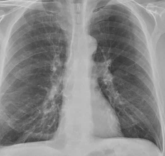
Thoracic spine x-rays provide a picture of the bones in the thoracic region. These include the ribs, thoracic spine, thoracic facet joints, and costovertebral joints. The heart shadow and lung fields are also well-visualized.
Benefits of thoracic spine x-rays include:
- Inexpensive
- Readily available
- Quick
- Painless
Risks associated with thoracic spine X-rays include:
- Radiation exposure.
X-rays are a form of energy. X-ray machines pass radiation through specific body parts to produce images. These images are called x-rays. The amount of radiation exposure depends upon what part of the body is being evaluated. For example, a single chest x-ray exposes a patient to about 0.1mSv (1). This is equivalent of a single day’s background radiation (2).
Common Spinal Conditions That May Require An X-Ray
Thoracic spine pain that is unresponsive to conservative care and medication requires further evaluation. A thoracic spine x-ray is an appropriate next step. There are many different causes of thoracic pain that can be identified on the x-ray. These include:
- Congenital Spinal Conditions
- Abnormal Spinal Curves
- Broken Bones
- Injury to the Spinal Disc
- Arthritis
- Osteoporosis
- Infections
- Scoliosis
- Kyphosis
Risks and Considerations
The principal risk of thoracic spine x-ray is radiation exposure in pregnant women. Exposure to high-dose radiation may affect the unborn child (3).
While important, thoracic spine x-rays do have limitations. They are not able to fully evaluate many structures which may be causing pain in the thoracic spine. These include.
- Thoracic Discs: a key shock absorber
- Thoracic Facet Joints: paired joints that limit rotation and provide stability.
- Costovertebral Joints
- Ligaments: duct tape that holds bones together is susceptible to injury creating instability
- Muscles: may be strained, torn, or ruptured
- Nerves: can be irritated, compressed, or cut
All the above structures can be well visualized and evaluated by a thoracic MRI. This is discussed in a different post.
X-Ray Imaging Procedure And Safety Protocols
- Section to discuss how the imaging works, what is involved in the process, and the safety protocols to ensure a safe process.
An x-ray machine is like a large camera that takes pictures of a specific body area. One example would be the thoracic spine. An x-ray machine produces a very concentrated beam of electrons known as x-ray photons. The x-ray photons can not be seen or felt. The beam travels through the body to create an x-ray picture. Dense materials in our bodies such as bones absorb the beam of electrons. Soft tissues such as skin and body organs are not able to absorb the beam of electrons. The result is an x-ray film. The black areas on an x-ray represent areas where the beam of electrons has passed through the soft tissue.
Safety Protocols
X-ray equipment should be maintained and operated by qualified staff and periodically tested. Technicians should wear protective eyewear and stand behind a lead-lined protective wall. All female patients are specifically asked whether they are pregnant or not.
Preparing For An X-Ray Exam
There are no fluid or medication restrictions. Avoid wearing jewelry. Tell the x-ray tech if you have a spinal cord stimulator or pacemaker.
During The Exam: Thoracic Spine X-Ray Positioning
There are 3 different x-ray views of the thoracic spine that are often requested.
A/P X-ray
This is the most common and requires the patient to lie on their back. The x-ray beams pass from front to back. Hence they are referred to as anterior-posterior x-rays.
Lateral X-ray
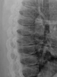
This is a side view of the thoracic spine and that requires the patient to lay on their side.
Oblique X-ray
This x-ray views the thoracic spine from an oblique view. It is beneficial in the evaluation of axillary ribs, thoracic facet joints, and pneumothorax (2).
After An X-Ray Procedure
Patients are allowed to leave the facility after completion of the thoracic x-ray with no fluid, food, or medication restrictions. Results are typically available within 24-48 hours.
Interpreting The X-Ray Results
After completion of the thoracic spine x-ray, a radiologist reviews the images. They examine the x-ray in a systematic fashion looking for abnormalities in the spine, ribs, facet joints, costovertebral joints, lung, and heart.
What Abnormalities In Results Could Mean?
Thoracic spine x-rays are powerful diagnostic tools as they can identify bone and some heart and lung abnormalities. The most common abnormalities include:
- Thoracic Disc Degeneration: Disc heights are narrowed.
- Bone Spurs: boney growth formations of varying size indicative of instability
- Spondylolisthesis: abnormal slippage of the vertebral body due to fracture.
- Thoracic Facet Overgrowth
- Rib Dislocation
- Rib Fracture
- Pneumonia
- Enlargement of the heart: Also referred to as cardiomegaly.
- Spinal Stenosis: narrowing of the spinal canal.
Get The Proper Diagnosis For Your Spinal Condition
Thoracic spine x-rays are a powerful diagnostic tool. They are readily available, quick, and inexpensive. Radiation is involved and women who are pregnant should avoid them. There are three common thoracic spine x-ray views: A/P, lateral, and oblique. Thoracic spine x-rays can identify abnormalizes in the spine, ribs, heart, and lungs.
At Centeno-Schultz Clinic we are experts in the diagnosis and treatment of thoracic injuries. We use a number of available tools including thoracic spine x-rays to make an accurate diagnosis and treatment plan. All physicians are board-certified and fellowship-trained. We will review your history, treatment to date, and radiographic studies within the office or from the comfort of your home or cabin. We will identify the most likely cause of your thoracic pain and your candidacy for regenerative treatments.
If you or a family member is struggling with thoracic pain which has been unresponsive to conservative care please contact us. We are here to help you understand your spinal health. A thoracic spine x-ray or MRI may provide valuable information previously not seen. To schedule please contact Jen at 720-287-7196 or [email protected] or Vanessa at [email protected]. It is time to stop suffering and start living life to its fullest.
We are here to help you understand your spinal health.
References:
1. Gargani L, Picano E. The risk of cumulative radiation exposure in chest imaging and the advantage of bedside ultrasound. Crit Ultrasound J. 2015 Mar 28;7:4. doi: 10.1186/s13089-015-0020-x. PMID: 25883779; PMCID: PMC4392040.
2.Broder J. Imaging the Chest: The Chest Radiograph. Diagnostic Imaging for the Emergency Physician. 2011:185–296. doi: 10.1016/B978-1-4160-6113-7.10005-5. Epub 2011 Jul 28. PMCID: PMC8139021.
3.Tulay CM, Yaldız S, Bilge A. Oblique Chest X-Ray: An Alternative Way to Detect Pneumothorax. Ann Thorac Cardiovasc Surg. 2018 Jun 20;24(3):127-130. doi: 10.5761/atcs.oa.17-00220. Epub 2018 Mar 16. PMID: 29553087; PMCID: PMC6033527.