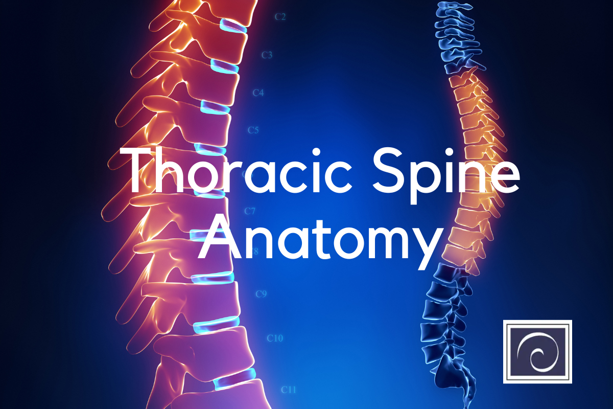
What Is The Thoracic Spine?
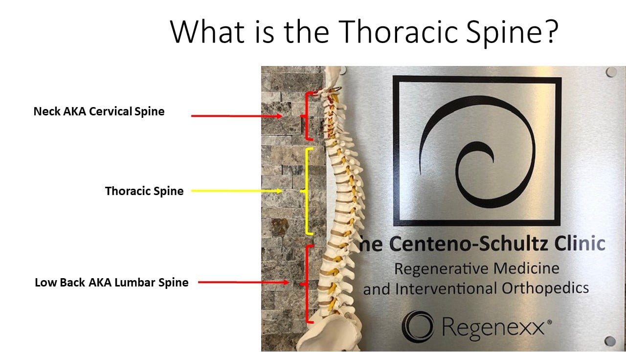
The thoracic spine refers to the section of the spinal column corresponding to the mid-back. It is between the cervical spine (neck) and the lumbar spine (low back). It consists of 12 boney building blocks called vertebral bodies that are labeled T1-T12. The T refers to the thoracic spine.
The thoracic spine is critical as it protects many vital structures, maintains the upright posture of the body, and provides multiple sites of attachment for muscles and ligaments. All this will be discussed in detail below.
Understanding The Vertebrae: T1-T12
The thoracic spine comprises 12 boney building blocks called vertebral bodies (1). They are stacked one upon another, much like bricks in a building. They are numbered from top to bottom: T1-12. Sandwiched between each vertebral body is an important cushion called a disc. The discs are labeled according to the two vertebral bodies they are sandwiched between.
For example, the first thoracic disc is referred to as the T1/2 disc as it is between the T1 and T2 vertebral bodies. Each disc has two principal components: a gelatinous center called the nucleus pulposus, and a thick supportive side wall called the annulus fibrous. Each is susceptible to injury and degeneration.
The thoracic spine has several unique features not found in the cervical or lumbar spine. The primary characteristic of the thoracic vertebrae is the presence of small joints formed where the rib connects with the vertebral body. These are called costal facet joints. The costal facet joints allow rotation, flexion, and rotation in the thoracic spine (2). At each level of the thoracic spine, there are 6 costal facets: 3 on the right and 3 on the left. They include:
Costotransverse Joint
A joint is formed where the rib connects with the transverse process of the vertebral body.
Costovertebral Joint
The rib connects at two points on each vertebral body per side. These are called demi facets. A costovertebral joint is formed where the rib connects with each demi facet. There are two costovertebral joints on the right and two on the left.
Other essential differences in the thoracic spine anatomy include:
The length of the transverse processes decreases as the column descends.
T5-T8 have the greatest rotation ability of the thoracic region.
T1, T10, T11, and T12 have complete costal facets.
The Structure And Function Of The Intervertebral Discs
Thoracic spine anatomy includes many important structures that can be injured, causing significant pain and dysfunction. The thoracic intervertebral disc is susceptible to injury and degeneration. Herniation of the intervertebral disc in the thoracic region makes up only 0.5% to 4.5%of all disc ruptures (3)
There are 12 intervertebral discs in the thoracic spine. The thoracic intervertebral discs are the fibrocartilaginous cushions located between the thoracic vertebrae in the spinal column. The discs are made up of two main components: the annulus fibrosus, which is the tough outer layer of the disc, and the nucleus pulposus, which is the soft, jelly-like center.
The annulus fibrosus is made up of layers of collagen fibers that are arranged in a crosshatch pattern, providing strength and stability to the disc. The nucleus pulposus is a gel-like substance that serves as a shock absorber, distributing pressure evenly across the disc during movement.
The function of the thoracic intervertebral discs is to provide support, flexibility, and cushioning to the spine (4). They act as shock absorbers, protecting the spinal column from the impact of daily activities such as walking, jumping, and running. The discs also allow for movement and flexibility in the spine, allowing the torso to bend and twist. As we age, the discs can become worn or damaged, leading to conditions such as herniated discs or degenerative disc disease.
Thoracic Spine Joints
A joint is where two or more bones meet to allow movement. The spinal column is made up of a series of these joints, which allow for flexibility and movement in the spine. There are two main types of intervertebral joints in the spine:
Cartilaginous Joints
A cartilaginous joint is where the bones are joined by fibrocartilage or hyaline cartilage. The joints between the vertebral bodies and the intervertebral disc are cartilaginous joints. They provide stability and support to the spine.
Synovial Joints
Synovial joints are the most common type of joint in the body. They join bones with a thick fibrous capsule. Unlike a cartilaginous joint, they have a joint cavity and a lubricant called synovial fluid. Synovial joints allow for movement and flexibility.
Synovial joints in the thoracic spine include:
Thoracic Facet Joint
A small, paired joint in the spinal column is located at the back of each thoracic vertebra. There is a right and left thoracic facet joint at each spine level. They provide stability and control the movement of the spine. They can become injured, resulting in pain.
Costotransverse Joint
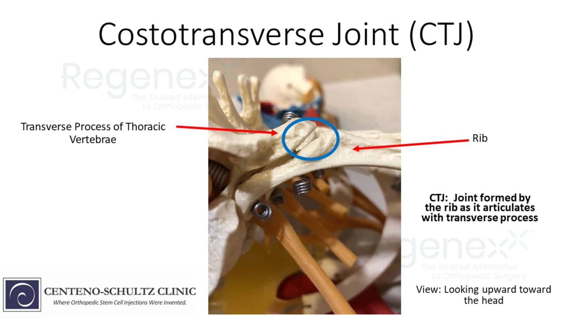
A small joint where the rib meets the transverse process of the vertebral body. The joint is formed by the articulation of the tubercle of the rib, a small bump on the back of the rib, and the transverse process, a bony projection on the side of the vertebra.
There are twelve pairs of costotransverse joints in the thoracic spine, one for each of the twelve ribs that attach to the thoracic vertebrae. The costotransverse joint allows for movement and flexibility in the thoracic spine, specifically in the area where the rib attaches to the vertebra
Costovertebral Joint
The costovertebral joint is a small joint where the rib meets the vertebral body. There are twelve pairs of costovertebral joints in the human body. The joint allows for movement and flexibility in the ribcage, specifically in the area where the rib attaches to the thoracic vertebra, while also providing stability and support to the spine and ribcage. The costovertebral joint is unique to the thoracic spine and is not present in the cervical or thoracic spine.
Muscles Of The Thoracic Spine
The muscles in the thoracic spine are important for maintaining posture, stability, and mobility of the upper back. They are arranged in layers.
Superficial Back Muscles
These are the muscles that are closest to the skin. They are large muscles that include:
Trapezius
Large triangular-shaped muscle that extends from the base of the skull to the middle of the back.
It is responsible for moving the shoulder blades and supporting the weight of the arms.
Rhomboids
Located between the shoulder blades and attach to the spine and the shoulder blades, the rhomboids help to stabilize the shoulder blades and assist in movements such as pulling the shoulder blades back and down.
Intermediate Back Muscles
These muscles are located deep in the superficial back muscles. As such they make up the middle layer of muscles in the thoracic spine. The erector spinae muscles are a group of muscles that run parallel to the spine and extend from the base of the skull to the sacrum. They help to maintain posture and assist in movements such as bending and twisting.
Deep Back Muscles
These muscles are located deep in the erector spinal muscles. They are a group of short muscles collectively referred to as transversospinales.
Thoracic Spine Ligaments
Ligaments are thick pieces of connective tissue that connect bone to bone. The ligaments of the thoracic spine play an important role in providing stability and support to the spinal column. The thoracic spine is the longest and most rigid section of the spine, and the ligaments help to keep the vertebrae in proper alignment and prevent excessive movement.
Here are some of the major ligaments in the thoracic spine.
Radiate Ligament
This ligament connects the rib head to the thoracic vertebrae (6). They have 3 bands: superior, middle, and inferior.
Costotransverse Ligament
This ligament connects the rib with the transverse process of the thoracic vertebrae. It supports and prevents dislocation of the ribs.
Lateral Costotransverse Ligament
This ligament connects the rib to the transverse process of the thoracic vertebrae.
Superior Costotransverse Ligament
This ligament connects the top portion of the rib to the transverse process of the thoracic vertebrae above.
Anterior Longitudinal Ligament
Important ligament extends across the front of the entire spine, providing support and stability for the vertebral bodies and disc.
Posterior Longitudinal Ligament
The counterpart of the anterior longitudinal ligament, it runs down the backside of the vertebral bodies. It helps to hold the vertebral bodies in place and prevent them from sliding backward.
Ligamentum Flavum
This ligament connects the lamina from C2/3 down to the sacrum.
Supraspinatus Ligament
This ligament connects the tips of the spinous processes in the cervical, thoracic, and lumbar spine.
Interspinous Ligament
Small pieces of connective tissue connect the mid portion of the spinous processes in the cervical, thoracic, and lumbar spine. They are stabilizers of the spine and limit spine flexion (7).
Thoracic Spine Nerves And Nerve Roots

The thoracic spine protects the spinal cord and nerve roots. The nerves and nerve roots of the thoracic spine are responsible for sending and receiving signals from the muscles, skin, and organs in the chest and upper abdomen. Important nerves in the thoracic spine include:
Thoracic Nerve Roots
Twelve pairs of thoracic nerve roots (T1-T12) emerge from the spinal cord between the thoracic vertebrae. These nerve roots join together to form the thoracic nerves.
Thoracic Nerves
The thoracic nerves supply the muscles and skin of the chest and upper abdomen. Each thoracic nerve runs a course along the corresponding thoracic vertebra and branches out to provide electrical information to specific muscles and areas of skin.
Sympathetic Nerves
The sympathetic nervous system is a branch of the autonomic nervous system that controls involuntary functions such as heart rate, blood pressure, and digestion. The sympathetic nerves pass through the thoracic spine and connect to the sympathetic ganglia.
Vagus Nerve
The vagus nerve is the tenth cranial nerve and plays a crucial role in regulating many vital functions of the body, including heart rate, breathing, and digestion. It passes through the thoracic spine and innervates organs such as the heart, lungs, and stomach.
Phrenic Nerve
The phrenic nerve originates from the cervical spine but passes through the thoracic spine and innervates the diaphragm, which is the main muscle involved in breathing.
Intercostal Nerve
Also known as ventral rami of the thoracic spinal nerves, these nerves originate from the front aspect of the spinal cord. The intercostal nerves run between the ribs and provide information to the muscles of respiration (intercostal muscles)
Dorsal Rami Of Thoracic Spinal Nerve
Small branches of the spinal nerves originate from the backside of the spinal cord. Dorsal rami of the thoracic nerves provide information to the muscles, joints, and skin of the thoracic region. They are responsible for the sensation of skin and movement of the thoracic muscles.
Thoracic Spine Range Of Motion – Normal Vs. Abnormal
The thoracic spine has a restricted range of motion during flexion and extension compared to the cervical and lumbar spine. The restricted range of motion is due to the ribs, which provide stability and protection to the thoracic organs. Thoracic spine mobility decreased with increasing age. (8). The thoracic spine has several different types of movements that include:
- Flexion: forward bending of the spine. The range of thoracic flexion is limited to 20-35 degrees.
- Extension: backward bending of the spine. The range of thoracic extension is between 20-40 degrees. Spinal mobilization can significantly improve thoracic extension (9).
- Lateral flexion: AKA side bending of the spine. The range of motion for thoracic lateral flexion ranges from 13-35 degrees in each direction (10)
- Rotation: Turning of the spine. The normal range of motion for thoracic rotation is limited to about 35-45 degrees (11).
Thoracic Spine Injury And Pathology
The thoracic spine, which consists of 12 vertebrae, is an essential part of the skeletal system. It provides stability and support for the upper body and involves various movements such as bending, twisting, and extending. Like the cervical and lumbar spine, the thoracic spine is susceptible to injury with resultant pain, restriction in range of motion, and dysfunction. The most common injuries and conditions include:
Herniated Thoracic Disc
A herniated thoracic disc is especially difficult because there are not as many treatments available as there are for disc herniations in other areas of the spine. To understand Thoracic Disc Herniations, though, we first need to cover thoracic spine anatomy and function. With disc herniation, the annulus fibrosus get small tears throughout the annulus. An annulus is a bunch of concentric fibers, so, as the fibers get damaged and cut, the pressure that is built up within the nucleus pushes the now weakened annulus outward, creating a bulge or herniation. The disc begins to weaken via mild degeneration/tearing of the annular fibers…
Read More About Herniated Thoracic DiscSpinal Stenosis
Spinal stenosis is the narrowing of the central spinal canal and is a cause of significant pain and disability. Common causes of spinal stenosis include disc protrusion, facet overgrowth and ligamentum flavum thickening. Surgery is often chosen when conservative therapies fail despite the lack of convincing evidence that it is a superior treatment option. Are there alternatives to back surgery for spinal stenosis? Yes. Regenexx DDD utilizes precise platelet injections into the facets, muscles, and ligaments to treat the lumbar stenosis, treating all of the components of the issue, which is crucial. Spinal stenosis is often an age-related condition attributed…
Read More About Spinal StenosisSpondylolisthesis
Spondylolisthesis means that one vertebra is slipping forward or backwards on another. This causes the hole where the nerve exits (foramen) to get smaller (also called foraminal stenosis). It also causes more wear and tear on the facet joint which can lead to arthritis or what’s called “facet hypertrophy”. spondylolisthesis recovery The amount of slippage is graded 1-4, with grade 1 meaning that the one vertebra has slipped up to 25% on the other vertebra. Grade 2 means that one bone has slipped from 25-50% with higher grades indicating more slippage. The vast majority of patients are grade 1 to 2.
Read More About SpondylolisthesisTorn Discs
The spinal discs are shock absorbers that live at each level between the vertebral bones (1). They have a tough outer annulus part and a soft inner gel part (nucleus pulposis). The outer covering can get damaged which can sometimes be seen on MRI and other times requires additional testing to identify. These tears are called: a torn disc, a disc tear, an annular tear, and when seen on MRI a “High-Intensity Zone” or HIZ. They can cause pain, mostly through ingrown nerves. There are torn disc findings that can be seen on MRI (HIZ) and these can be either asymptomatic (i.e. not painful) or…
Read More About Torn DiscsDegenerative Scoliosis
Degenerative Scoliosis, also known as Adult-onset Scoliosis, is a medical condition that involves a side bending in the spine. The bending can be mild, moderate, or severe with side-bending to either the right or the left. The term degenerative means generalized wear and tear and is common as we get older. Degenerative scoliosis is the curvature of the spine that occurs as a result of degeneration of the discs, small joints, and building blocks. The Degenerative Scoliosis curve is often located in the low back and forms a ‘C” shape. There is a convex and a concave side. The convex side is the open side where it curves outward.
Read More About Degenerative ScoliosisMake Educated Decisions About Your Thoracic Spine Treatment
The thoracic spine refers to the section of the spinal column corresponding to the mid-back. It comprises many dynamic and interrelated parts that include vertebral bodies, intervertebral discs, thoracic facet joints, costotransverse joints, costovertebral joints, muscles, ligaments, and nerves.
All these structures are susceptible to injury and degeneration with resultant pain and limitation. The thoracic spine is unique in that it has ribs that form joints at each level and provide a protective shell for many vital organs, such as the lungs and heart. The thoracic spine is complex and requires years of dedicated study. Thoracic pain is all too often ignored or misdiagnosed.
The Centeno-Schultz Clinic is an expert in the evaluation and treatment of thoracic spine injuries. A board-certified, fellowship-trained physician can review your current thoracic complaint, past medical history, past surgical history, and radiographic imaging and determine whether you are a candidate for regenerative therapies. If your thoracic pain has failed to respond to conservative treatment and is limiting your life, please consult us today. It is time to stop the suffering.
Experiencing pain in your thoracic spine? Consult with us today.
1.Gkasdaris G, Tripsianis G, Kotopoulos K, Kapetanakis S. Clinical anatomy and significance of the thoracic intervertebral foramen: A cadaveric study and review of the literature. J Craniovertebr Junction Spine. 2016 Oct-Dec;7(4):228-235. doi: 10.4103/0974-8237.193266. PMID: 27891032; PMCID: PMC5111324.
2.Waxenbaum JA, Reddy V, Futterman B. Anatomy, Back, Thoracic Vertebrae. 2022 Aug 8. In: StatPearls [Internet]. Treasure Island (FL): StatPearls Publishing; 2023 Jan–. PMID: 29083651.
3.Göçmen S, Çolak A, Mutlu B, Asan A. Is back pain a diagnostic problem in clinical practices? A rare case report. Agri. 2015;27(3):163-5. doi: 10.5505/agri.2015.69782. PMID: 26356107.
4.Adams, Michael A. PhD*; Roughley, Peter J. PhD†. What is Intervertebral Disc Degeneration, and What Causes It?. Spine 31(18):p 2151-2161, August 15, 2006. | DOI: 10.1097/01.brs.0000231761.73859.2c
5.Göçmen S, Çolak A, Mutlu B, Asan A. Is back pain a diagnostic problem in clinical practices? A rare case report. Agri. 2015;27(3):163-5. doi: 10.5505/agri.2015.69782. PMID: 26356107.
6.Saker E, Graham RA, Nicholas R, D’Antoni AV, Loukas M, Oskouian RJ, Tubbs RS. Ligaments of the Costovertebral Joints including Biomechanics, Innervations, and Clinical Applications: A Comprehensive Review with Application to Approaches to the Thoracic Spine. Cureus. 2016 Nov 11;8(11):e874. doi: 10.7759/cureus.874. PMID: 27994992; PMCID: PMC5154401.
7.Iwanaga J, Simonds E, Yilmaz E, Schumacher M, Patel M, Tubbs RS. Anatomical and Biomechanical Study of the Lumbar Interspinous Ligament. Asian J Neurosurg. 2019 Nov 25;14(4):1203-1206. doi: 10.4103/ajns.AJNS_87_19. PMID: 31903363; PMCID: PMC6896651.
8. Kuo YL, Tully EA, Galea MP. Video based measurement of sagittal range of spinal motion in young and older adults. Man Ther. 2009 Dec;14(6):618-22. doi: 10.1016/j.math.2008.12.006. Epub 2009 Feb 6. PMID: 19201248.
9. Hwangbo G, Lee CW, Kim SG. Effects of spinal mobilization on range of motion and clinical outcomes in patients with chronic low back pain: a randomized controlled trial. Journal of Manipulative and Physiological Therapeutics. 2013 Nov-Dec;36(9):584-9. doi: 10.1016/j.jmpt.2013.07.005.
10. Budziński Ł, Białoszewski D, Koczy B, Wozniewski M. Effects of a stretching program on thoracic spine mobility in healthy young adults: a randomized controlled trial. Journal of Back and Musculoskeletal Rehabilitation. 2015;28(1):149-55. doi: 10.3233/BMR-140523.
11. Nee RJ, Butler DS. Management of peripheral neuropathic pain: integrating neurobiology, neurodynamics, and clinical evidence. Physical therapy in sport. 2012 Aug 1;13(3):213-22. doi: 10.1016/j.math.2011.10.005.