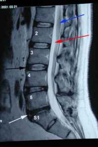
Blood supply to a given structure provides essential nutrients for growth, maintenance and repair. Some structures such as the heart have a rich blood supply with multiple arteries. Other structures have a very limited, tenuous blood supply which places them at risk for impaired growth, repair and potential cellular death. The lumbar intervertebral disc is such a structure. While it is the cornerstone of our spine and bears the weight of our bodies as we walk, its blood supply is extremely limited.
What does this mean? If injuries occur the lumbar disc has limited capability of repairing itself due to its very limited blood supply. The result is a slow insidious degeneration of the lumbar disc characterized by reduction in disc height and signal. The MRI below on the left illustrates a degenerative L5/S1 disc. The blue arrow identifies the spinal cord, the red arrow is pointing to the cerebral spinal fluid and the black arrow identifies the L3/4 disc. Note the L3/4 disc has a white signal within the disc which represents hydration. It also is identical in height and brightness to the disc above it. Both of these discs are normal. The white arrow identifies a degenerative L5/S1 disc which is black in color with reduction in height in comparison to the adjacent discs.

With injury there is also a propensity to develop bulges since the integrity of the side wall (annulus fibrosis) is compromised.

If sufficient damage occurs, a disc bulge can progresses to a disc herniation where an portion of the inner contents of the disc (nucleus pulposus) are extruded.

In a previous blog I discussed the importance of platelets and the four major growth factors they contain: Platelet-derived growth factor (PDGF), transforming growth factor beta (TGF-b), vascular endothelial growth factor (VEGF) and epithelial growth factor (EGF). Vascular endothelial factor is responsible for angiogenesis (the creation of new blood vessels). These new blood vessels can provide essential nutrients to repair damaged tissue. At Regenexx we injected concentrated VEGF adjacent a degenerative disc with the hope of improving blood flow and initiating repair. Please see MRI results below. On the left are MRI images of the L5/S1 disc prior to therapy. Note there is a reduction in height and brightness of the L5/S1 disc. On the right are MRI images of the same L5/S1 disc after therapy. Note an increase in the height and the signal of the disc. There is significant improvement in the disc height and signal. What is the significance? This is a patient who despite prior back surgery continued to have pain. After therapy at Regenexx the patient had near complete resolution of pain as well as MRI evidence of lumbar disc repair.
