The headaches, neck pain and dizziness are debilitating and getting worse. Conservative treatments and medications have failed. Your doctor has referrred you to a neurosurgeon. He mentioned Chiari Decompression surgery. What is a Chiari Malformation? What are the three different types of Chiari Malformations? What are the symptoms of Chiari Malformation? What is Chiari Decompression surgery? What are the 8 major Chiari Decompression surgery complications? What are the long-term effects of Chiari Decompression surgery? Let’s dig in.
What Is a Chiari Malformation?
A Chiari Malformation is a medical condition where a part of the brain at the back of the skull abnormally descends through an opening in the skull. It is named after Dr. Hans Chiari who was an Austrian pathologist who in the late 1880’s studied deformities of the brain.
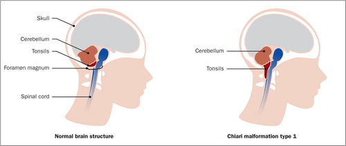
The brain is a large structure divided into different parts that reside within the skull. Important parts of the brain called the Cerebellum and Brainstem sit at the base of the skull. The Foramen Magnum is a large hole at the base of the skull that allows the brain to join the spinal canal. The Cerebellum and Brainstem under normal conditions sit above the boney opening at the base of the brain (Foramen Magnum).
In Chiari Malformations the cerebellum and brainstem move below the level of Foreman Magnum putting pressure on the lower parts of the brain (1). It can also affect the flow of spinal fluid around the brain and spinal cord.
Chiari Malformation can occur when the opening at the base of the skull is not large enough or when there is abnormal pressure in the brain pushing it downward.
Are there Different Types of Chiari Malformations?
Yes! Chiari Malformations are categorized into 4 major types based upon anatomical abnormality and whether congenital defects are present (2).
Chiari Type 1 Malformation
This malformation occurs during fetal development and is characterized by downward movement of the Cerebellum beneath the boney opening at the base of the skull (Foramen Magnum). It is typically 4 mm. That means that the Cerebellum and Brainstem descent 4mm beneath the Foramen Magnum. Why is this important?
It can block the flow of spinal fluid between the spinal canal and brain leading to an increase in pressure. It can also put pressure on the lower portion of the brain with varying symptoms. Chiari Type 1 Malformation is typically associated a fluid-filled cyst within the spinal cord which is called a Syrinx.
Chiari Type II Malformation
In Chiari Type II Malformation a greater amount of tissue extends down into the spinal canal compared to Chiari Type 1. It is usually associated with a Myelomeningocele which is a form of Spina Bifida. This is a condition where the spinal canal and the backbone do not close before birth.
Chiari Type III Malformation
This is the most severe form of Chiari Malformation. A significant amount of the brain is pushed through the opening at the base of the skull. This form of CM is diagnosed at birth or with ultrasound during pregnancy.
Chiari Type IV Malformation
Type IV is rare and involves an incomplete or underdeveloped Cerebellum. This is also referred to as Cerebellar Hypoplasia. In this type of CM the Cerebellum is in its normal position in relation to the Foramen Magnum but parts of it are missing.
Symptoms of a Chiari Malformation
Many people with Chiari Malformation have no signs or symptoms (3). Symptoms can change depending upon the amount of brain and nerve compression and increase in Cerebral Spinal Fluid pressure. The downward displaced Cerebellum and Brainstem restrict the flow of Cerebral Spinal Fluid from the brain into the spinal cord. Accordingly, it backs up with increased pressure within the skull. Abnormal pressure is also applied to the Cerebellum and Brainstem. The Cerebellum controls the coordination of movement. You know the wonderful coordination that is involved with walking, talking, dancing, and running. The Brainstem on the other hand controls many basic life functions such as heart rate, blood pressure, breathing, and level of consciousness.
Symptoms may include:
- Headache
- Neck Pain
- Dizziness
- Ringing in the ears (tinnitus)
- Arm and Leg weakness and numbness
- Difficulty with swallowing
- Breathing problems
- Problems with hand coordination
- Nausea and Vomiting
- Balance problems
What Is Chiari Decompression Surgery?
Chiari Decompression surgery can also called a Posterior Fossa Decompression or Craniectomy. The Cranium is that part of ther skull that contains the brain. Chiari Decompression surgery involves removing a bone at the base of the skull so as to widen the boney opening at the base of the skull. This opening is called the Foramen Magnum.
The Decompression surgery has four principal goals:
- Create more space for the brain
- Slow or stop the progression of symptoms
- Relieve compression of the Cerebellum and Brainstem
- Restore normal cerebral Spinal Fluid flow
Chiari Decompression surgery can also involve additional surgical procedures depending upon the severity of the underlying condition and a patient’s symptoms. These include:
Dural Patch
The Dura is one of three layers of connective tissue that cover and protect the brain and spinal cord. A patch is sewn into the place so as to expand the space. It is akin to letting out the waistband in a pair of pants that are too tight.
Laminectomy
The Lamina are flat bones located on the backside of the spine. They form the roof of the spinal canal and are present at each level of the spine. The Lamina protects the spinal cord, spinal nerves and spinal fluid. Laminectomy is the surgical procedure where a portion of the Lamina is removed. A Laminectomy is typically performed to gain access to the spinal cord and relieve pressure on the spinal cord and nerves.
Fusion
Fusion is a surgical procedure where one of more spinal levels are mended together using surgical screws and plates. For example a C2-C3 Fusion where the joint is welded together using plates and screws. The previously mobile joint becomes immobile and ” more stable” as a result of the Fusion surgery.
Who Is a Candidate for Chiari Decompression Surgery?
Surgical recommendations can vary depending upon a surgeon’s training, experience and a given patient’s clinical presentation. In general Chiari Decompression Surgery is indicated for patients with Chiari type 1 Malformations that are symptomatic (4).
What Are the Long-Term Effects of Chiari Decompression Surgery and It’s Complications?
The long-terms effects of Chiari Decompression Surgery depend many factors that include the severity of the underlying condition, a patient’s symptoms, prior medical and surgical history and the exact type of surgery performed. The most common complications from Chiari Decompresssion surgery include:
Infection
Infection may be limited to skiin or extend into bone and neural tissue including the brain and spinal cord. The later is known as Meningitis and is a serious medical condition (5). Antibiotics can help in some cases whereas additional surgery is often required.
No Clinical Improvement
Surgery is performed to improve a patient’s condition and well being. Unfortunately there is no guarantee or assurance that is will occur. Some patients who undergo Chiari Decompression surgery have no clinical benefit. This can be devastating to patients and families alike.
Nerve Injury
Removal of the bone as the base of the skull (decompression) requires retraction and tension to be placed on the Occipital and Cranial nerves. This can result in temporary or permanet injury to the nerves. Patients can suffer from new onset upper neck and head pain. Ouch!
Spinal Instability
Stability of the upper neck is dependent upon muscle strength, tendon and ligament stability. The Subocciptial muscles are group of four muscles located at the base of the skull that provide important stabilization of the upper neck. They attach onto or near the bone that is surgically removed during Chiari Decompression. Surgery can compromise, injury or weaken these important stabilizing muscles and tendons. Ligaments are thick pieces of connective tissue that connect bone to bone. In many cases these ligaments are cut or damaged during Decompression surgery leading to upper neck instability.
Pseudomeningocele
A Pseudomeningocele is a known complication after spinal surgery (6). It is an abnormal collection of Cerebrospinal fluid that occurs outside of the spinal canal. Said another way it is a pocket of spinal fluid that occurs in the soft tissue of the neck. It occurs as a result of a leak in one of the coverings of the spinal cord. Spinal fluid flows out of the spinal canal and collects in the soft tissue. Treatment often times requires additional surgery and patching the leak.
Cerebral Spinal Fluid Leak
The Dura is a layer of connective tissue that surrounds and protects the brain and spinal cord. Decompression surgery can injure this protective layer resulting in a leak of spinal fluid. The symptoms of such a leak include a severe headache that is made worse when being upright, nausea, vomiting, neck pain, changes in hearing and sensitivity to light.
Failed Fusion
A failed Fusion occurs when the bones that are brought together surgically using screws, plates and bolts fail to unite. This is a significant problem as it can cause significant upper neck instability and pain. Treatment requires additional surgery often times with extension of the Fusion.
Increased Pain and Dysfunction
Removal of the base of the skull bone, cutting of important ligaments, traction on nerves, muscles, and Dura can result in an increase in pain and dysfunction. Surgery and removal of bones is permanent and changes the biomechanics of the upper neck forever.
In Conclusion
A Chiari Malformation is a medical condition where a portion of the brain tissue protrudes into the spinal canal through the Foramen Magnum.
Chiari Malformations are categorized into 4 major types based upon anatomical abnormality and whether congenital defects are present.
There are 4 major types of Chiari Malformations.
Many people with Chiari Malformation have no signs or symptoms.
Common symptoms of Chiari Malformations include headache, neck pain, dizziness, nausea and vomiting.
Chiari Decompression surgery is recommended for patients with Chiari type 1 Malformations that are symptomatic.
Chiari Decompression surgery involves removing a bone at the base of the skull to create more space for the brain and spinal cord.
Chiari Decompression surgery may also involve additional surgeries such as a Dural Patch, Laminectomy and Cervical Fusion.
The 8 major complications associated with Chiari Decompression surgery are:
- Infection
- No Clinical Improvement
- Nerve Injury
- Spinal Instability
- Pseudomeningocele
- Cerebral Spinal Fluid Leak
- Failed Fusion
- Increased Pain and Dysfunction
The Centeno-Schultz Clinic are experts in the evaluation and treatment of neck pain, headaches and associated neurologic symptoms. Headache, dizziness, and neck can be caused by other conditions other than Chiari Malformation. We are committed to securing an accurate diagnosis so that the best and appropriate treatment plan can be started. Treatment options include precise image guided injection procedures of PRP and Bone Marrow Concentrate which contains stem cells, into injured joints, ligaments and supporting tissue. Many cases of upper neck pain and headache are due to instability of the upper spine. This is called Craniocervical Instabilty (CCI). To learn more about CCI please click on the video below.
Surgery is permanent. There are no refunds. The landscape and mechanics of the neck are changed forever.
If you or a loved one have ongoing neck, headache, and dizziness that has not responded to conservative treatment, please schedule a telephone candidacy discussion with a board-certified, fellowship-trained physician. At the Centeno-Schultz Clinic, we are experts in the evaluation and treatment of upper neck injuries. From the comfort of your home or office learn what treatment options are available for you.
1.Fernández AA, Guerrero AI, Martínez MI, et al. Malformations of the craniocervical junction (Chiari type I and syringomyelia: classification, diagnosis and treatment). BMC Musculoskelet Disord. 2009;10 Suppl 1(Suppl 1):S1. Published 2009 Dec 17. doi:10.1186/1471-2474-10-S1-S1
2,2.Shah AH, Dhar A, Elsanafiry MSM, Goel A. Chiari malformation: Has the dilemma ended?. J Craniovertebr Junction Spine. 2017;8(4):297-304. doi:10.4103/jcvjs.JCVJS_138_17
3,Abd-El-Barr MM, Strong CI, Groff MW. Chiari malformations: diagnosis, treatments and failures. J Neurosurg Sci. 2014 Dec;58(4):215-21. PMID: 25418275.
4,Giammattei L, Borsotti F, Parker F, Messerer M. Chiari I malformation: surgical technique, indications and limits. Acta Neurochir (Wien). 2018 Jan;160(1):213-217. doi: 10.1007/s00701-017-3380-0. Epub 2017 Nov 12. PMID: 29130121.
5,Dubey A, Sung WS, Shaya M, Patwardhan R, Willis B, Smith D, Nanda A. Complications of posterior cranial fossa surgery–an institutional experience of 500 patients. Surg Neurol. 2009 Oct;72(4):369-75. doi: 10.1016/j.surneu.2009.04.001. Epub 2009 Jul 14. PMID: 19604553.
6. Weng YJ, Cheng CC, Li YY, Huang TJ, Hsu RW. Management of giant pseudomeningoceles after spinal surgery. BMC Musculoskelet Disord. 2010;11:53. Published 2010 Mar 21. doi:10.1186/1471-2474-11-53
Am I a Candidate?
To answer this question, fill out the candidate form below to request a new patient evaluation, and a patient advocate will reach out to you to determine your next steps. Your one-hour, in-office or telemedicine evaluation will be with one of the world’s experts in the field of Interventional Orthopedics.