We have developed an active community of patients with craniocervical instability who are following our PICL procedure. So what goes into performing this procedure? There’s quite a bit, so let’s dig in.
CCI and PICL
CCI stands for cranial cervical instability which means that the ligaments that hold the head on are too loose. To help that problem, we developed a new procedure called PICL which stands for Percutaneous Implantation of the CCJ Ligaments. This is still an investigational procedure that has already changed many lives and helped patients avoid a complication-laden upper cervical fusion. To learn more, see my video below:
What Goes into the PICL Procedure?
Given the interest in this procedure from patients who have CCI, there are lots of questions. Since the procedure isn’t yet covered by insurance, a few have asked why it costs more than their co-pay and deductible if they decide to have a more invasive cervical fusion. They have also asked why we don’t authorize any other Regenexx site to perform this procedure. The only way to answer that is to go behind the scenes to understand all of the things that go into a properly performed PICL procedure:
Development of the Procedure
Nobody had ever injected these ligaments before using x-ray guidance with contrast confirmation before I attempted it in 2014. These are the alar, accessory, and transverse ligaments. Hence, everything was new. In fact, we didn’t even know if these ligaments could be reached this way until the first injection.
We had to figure out:
- How to position the patient
- How to keep the area at the back of the throat being injected sterile
- How to keep the mouth open and tongue depressed with something that could be easily x-rayed through
- How to see the needle on the fluoroscope
- Where the actual ligaments lived in relationship to the bony landmarks
- How to prevent infection
- How to maximize access to various ligaments
This is just a shortlist.
Taking the Bone Marrow
Due to concerns about infection in this area, we moved to a full surgical prep and drape for the bone marrow aspiration, That technique is specialized as well, to maximize the number of stem cells in the sample, which takes longer than the average BMA performed at the average clinic using bone marrow concentrate.
Processing the Bone Marrow
This is a critical step and one of the biggest reasons why we will not allow any sites outside of our Colorado office to perform the PICL procedure. Colorado is unique in that it has a large c-GMP air-handing ISO-7 facility to process cells.
The bone marrow aspirate is then processed by highly trained and monitored technicians who are only permitted to work is ISO-5 sterile hoods, and there is constant surveillance for contamination in this environment which is a higher standard than the cleanest operating rooms. These technicians then produce high-dose bone marrow concentrate. The cost of this set-up? All-in, about a million USD to build and outfit a facility like this one.
Why is it needed? An infection in a PICL procedure due to contaminated bone marrow could be a life-threatening event.
Here is a public service announcement that stresses why you should be highly selective of where you get your PICL procedure:
Jump in on a Deep Dive of the PICL procedure with Alex Oxford and Dr. John Schultz
A Mouthpiece
The problem we had was that every mouthpiece we looked at that could keep the mouth open and tongue depressed to allow the procedure was metal and couldn’t be x-rayed through. We ultimately found one that was plastic and designed for GI use, but it was mediocre at best. We then set out to design our own and had many different iterations were 3D printed until deciding on two models in a few sizes each.
While one design worked better in patients with a certain mouth and facial anatomy, the other worked better in patients with different anatomy. One of the two is shown here. We’ve spent tens of thousands designing, prototyping, and producing these mouthpieces for our practice’s own use.
Cleaning and Visualizing the Area to be Injected
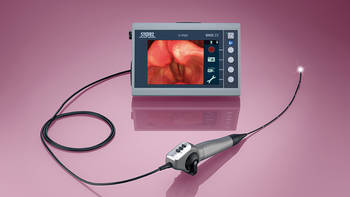
There’s really only one best practice way to “see” the back of the throat, where we place the needles in the PICL procedure, which is using endoscopy. This means that the flexible tube that is normally used by ENT surgeons is inserted into the mouth to visualize the injection site and clean it off.
Keeping the back of the throat clean is critical to avoid seeding bacteria into the upper neck ligament area, which could cause a meningitis-type infection. Endoscopes generally run about 40-70K.
Anesthesia
One of the challenges of the PICL procedure is that the patient is placed asleep using IV anesthesia, but the injection is occurring in the same space the patient uses for breathing (the airway). In addition, the patient’s throat must be kept dry. All of this is much more technically demanding than the average spinal injection.
Placing the Needles
This requires c-arm fluoroscopy and a deep understanding of the upper cervical anatomy. The fluoroscope itself requires an x-ray tech plus a lead shielded room. This machine costs about 130K and the room costs more.
Other Expertise
Many of these CCI patients also have upper neck joint damage and other ligaments involved. Take for example the C0-C1 and C1-C2 facet joints. Very few physicians understand how to perform these procedures safely and even fewer have done any significant number. In medicine, like anything else, frequency builds competency.
For C0-C1, I would estimate that we only have 100 US physicians who have done more than 20 of these injections.
Why?
The vertebral artery is close by and most physicians steer clear because you can’t visualize the opening of this joint on x-ray. If we up that number of required C0-C1 procedures to 100 per doctor, which would be the minimum number to begin to see higher levels of competency, we’re likely down to a handful of US or European physicians. How many have we done? I had my office manager pull the number of C0-C1 and C1-C2 procedures we have performed in approximately the last 5 years and the number came out to 2,000! It’s highly likely that there is no clinic on Earth that would even approach 200 in that time period.
In addition, we developed and published a new way to inject this joint (1).
The upshot? At the end of the day, there are many good reasons why the PICL procedure is only performed at our Broomfield, Colorado site. There are also reasons why it’s much more than a simple injection. I’ve actually only listed a few here.
See how the PICL procedure helped Bradley Gordon.
Conditions the PICL is Used to as Treatment
Atlantoaxial Instability (AAI)
Instability simply means that bones move around too much, usually due to damaged ligaments. In the spine, this can cause nerves to get banged into and joints to get damaged. In the craniocervical junction, instability can cause the upper cervical spinal nerves to get irritated, leading to headaches. In addition, the C0-C1 and C1-C2 facet joints can also get damaged. In addition, there are other nerves that exit the skull here that can get irritated, like the vagus nerve, which can cause rapid heart rate. What’s the Difference Between CCI and AAI? CCI refers to instability in any part of the craniocervical junction…
Read More About Atlantoaxial Instability (AAI)CCI
Craniocervical Instability is a medical condition characterized by injury and instability of the ligaments that hold your head onto the neck. Common symptoms of Cranial Cervical Instability include a painful, heavy head, headache, rapid heart rate, brain fog, neck pain, visual problems, dizziness, and chronic fatigue.CCI or neck ligament laxity treatment options depend upon the severity of the instability and clinical symptoms. When appropriate, conservative care should always be the first-line treatment. Craniocervical Instability Surgery is often recommended when conservative care fails. This involves a fusion of the head to the neck which is a major surgery that is associated with significant risks and complications…
Read More About CCICervical Medullary Syndrome
Cervical Medullary Syndrome is a clinical condition that occurs as a result of inflammation, deformity, or compression of the lower part of the brain. Symptoms can be extensive with fluctuating severity based upon the extent of the underlying injury. For example, mild irritation of the brainstem may cause only mild, intermittent symptoms. The upper cervical spine and brain are complex with multiple structures. These structures reside within the skull and protective confines of the cervical spine. Neither expands to accommodate inflammation, injury, and disease. Rather the delicate tissues of the brain and spinal cord are irritated or compressed. The 4 major conditions that cause cervical medullary syndrome are…
Read More About Cervical Medullary SyndromeChiari Malformation
Chiari Malformation Is a medical condition where a part of the brain at the back of the skull abnormally descends through an opening in the skull. It is named after Dr. Hans Chiari who was an Austrian pathologist who in the late 1880’s studied deformities of the brain.The brain is a large structure divided into different parts that reside within the skull. Important parts of the brain called the Cerebellum and Brainstem sit at the base of the skull. The Foramen Magnum is a large hole at the base of the skull that allows the brain to join the spinal canal. The Cerebellum…
Read More About Chiari MalformationCraniocervical Instability
Craniocervical Instability is a medical condition characterized by injury and instability of the ligaments that hold your head onto the neck. Common symptoms of Cranial Cervical Instability include a painful, heavy head, headache, rapid heart rate, brain fog, neck pain, visual problems, dizziness, and chronic fatigue.CCI or neck ligament laxity treatment options depend upon the severity of the instability and clinical symptoms. When appropriate, conservative care should always be the first-line treatment. Craniocervical Instability Surgery is often recommended when conservative care fails. This involves a fusion of the head to the neck which is a major surgery that is associated with significant risks and complications…
Read More About Craniocervical InstabilityReferences:
(1) Centeno C, Williams CJ, Markle J, Dodson E. A New Atlanto-Occipital (C0-C1) Joint Injection Technique. Pain Med. 2017 Oct 27. doi: 10.1093/pm/pnx256.
Am I a Candidate?
To answer this question, fill out the candidate form below to request a new patient evaluation, and a patient advocate will reach out to you to determine your next steps. Your one-hour, in-office or telemedicine evaluation will be with one of the world’s experts in the field of Interventional Orthopedics.


