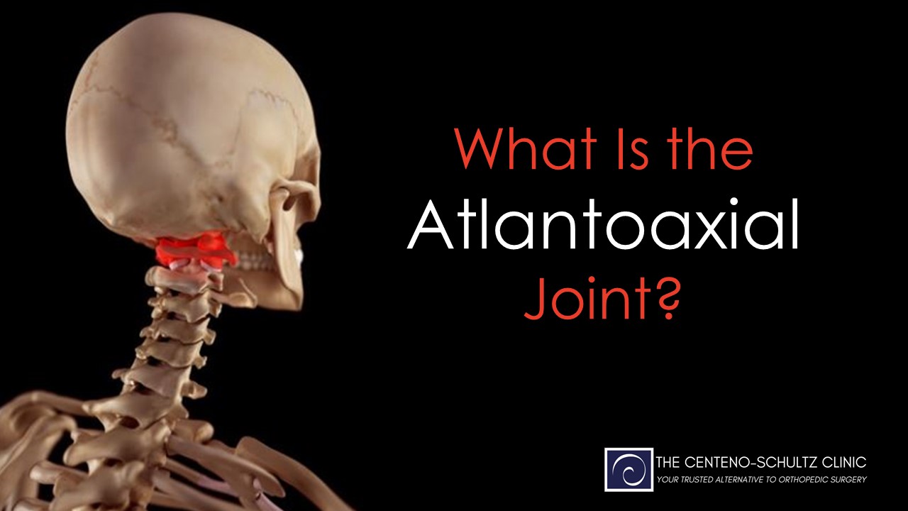
The headaches and upper neck pain are getting worse. Medication and therapy has not helped. Radiographic studies are normal. Your chiropractor thinks it may be coming from your upper neck. What is the Atlas? What is the Axis bone? What is the Atlantoaxial joint? What are the key components of the Atlantoaxial Joint? What is Atlantoaxial Joint’s prinicpal function? What is the Craniocervical Junction? Why is the Craniocervical Junction important?
Let’s dig in.
What Is the Atlas?
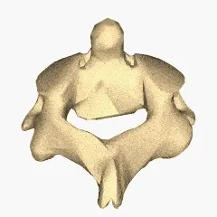
Atlas AKA C1 Bone
The Atlas bone, also known as the C1 is the first bone in your neck (1). It is named after the Greek God Atlas who held up the world on his shoulders. The Atlas plays an important role as your head rests directly on this bone.
What Is the Axis Bone
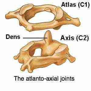
Axis AKA C2 Bone
The Axis bone is the second bone in the neck and as such it is also referred to as C2 (2). It is an odd-looking bone as it has a boney peg that looks like the Washington Monument. This boney projection is called the Dens. The Dens extend upward to the Atlas bone. It provides a stable point upon which the Atlas bone can rotate.
What Is the Atlantoaxial (AA) Joint?
The AA joint is formed by the union of the C1 and C2 bones. The Atlantoaxial joint is composed of three smaller joints:
Medial AA joint
This is a pivot type joint and is formed between the Dens and ring of the C1 bone.
Two Lateral AA joints
Bilateral joints formed by the C1 and C2 bones. A joint is formed when two bones come together. Think of where the thigh bone meets the shin bone. The two bones come together to form the knee joint. So too with the C1 and C2 bones. They come together and form a joint. Like the knee joint, the AA joint is lined with cartilage.
What Are the Key Components of the AA Joint?
Cartilage
Like the knee joint, the AA Joint is lined with cartilage. Cartilage is the white shiny surface on the end of a bone that enables the smooth, pain-free movement of the joint. It absorbs and distributes large compressive and shear forces. The AA Joint is susceptible to injury and degeneration resulting in pain and reduced range of motion.
Ligaments
Ligaments are thick pieces of connective tissue that connect bone to bone. Ligaments provide support and stability. There are several important ligaments which include:
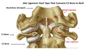
Alar Ligament
A key ligament that provides stability for the upper neck and head area. It essentially connects and stabilizes your head onto your neck. It connects the C2 bone to the skull.
Transverse Ligament
It forms a snug seatbelt around the Dens providing important stability for the C1/2 joint and upper cervical spine. It arches across the ring of the C1 bone holding the Dens in place as illustrated below.
Apical Ligament
A thin small ligament located at the top of the Dens bone and stretches upward to the base of the skull. It joins the Anterior Atlantoocciptal Membrane to provide support for the upper cervical spine.
Tectorial Membrane
The Tectorial Membrane is the superior continuation of the Posterior Longitudinal Ligament( PLL). It covers the backside surface of the Dens bone which is the bone that looks like the Washington Monument.
Capsule
The AA Joint has a capsule that covers the entire joint, It is a saran wrap-like covering composed of fibrous connective tissue. It provides support, distributes the forces, and limits the range of motion.
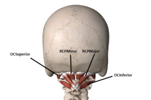
Muscles
The are a large number of small muscles in the upper neck. They provide important support, stability and enable motion such as bending forward, backwards and rotating. One of the muscles called the Rectus Capitus Posterior Minor (RCPM) plays a very important role. It actually connects to the covering of the brain (3). It is called the Myodural Bridge. Clinically physicians have found that a tight or dyfucntional Rectus Capitus Posterior Minor muscle can put pressure on the Dura resulting in severe headaches.
What Is the Function of the Atlantoaxial Joint?
The Atlantoaxial Joint is one of the most mobile joints in the spine. Rotation is the principal function. It allows us to turn our heads to the left and right. The AA joint enables us to move our heads in the familiar ” no” motion. It also allows limited flexion, extension, and lateral flexion.
The Atlantoaxial (AA) Joint is also part of the Craniocervical Junction. The Craniocervical Junction is the area between the Skull and the Cervical Spine (4). It consists of the bone that forms the base of the Skull, the first two bones in the Spine, and the neural structures that pass from the brain down into the Cervical Spine.
What Is In the Craniocervical Junction?
The base of the Skull also known as the Occiput has a large opening at its base called the Foramen Magnum (5). Foramen meanings opening. Magnum means large. So the Foramen Magnum is a large boney opening at the base of the Skull that allows important structures to pass through. Important structures that pass through the Foramen Magnum include:
Spinal Cord
The Spinal Cord consists of neural tissue that starts at the base of the brain and extends down into the low back. It is a cylindrical bundle of nerve fibers that control our voluntary and involuntary bodily functions. It carries signals between the brain and the rest of the body. As the Spinal Cord descends from the skull and through the neck and rest of the body it is protected on all sides by spinal bones. These bones provide boney armor to protect against injury. The Spinal Cord has an additional layer of protection afforded by the spinal fluid. The spinal fluid is also known as Cerebral Spinal fluid. It surrounds the Spinal Cord and extends the entire length of the spine. The image to the right is a side view of the Spinal Cord as it exits the brain. The Spinal Cord is black in color. The white that surrounds the Spinal Cord is the spinal fluid.
Cranial Nerves
As the Spinal cord descends through the Foramen Magnum and down the Spine important nerves branch off traveling to different parts of the body. There are a large number of nerves. These include the 12 Cranial nerves some of which control muscles whereas others are connected to internal organs such as the heart and lungs.
Arteries and Veins
Arteries and veins provide blood flow to and from important structures in the head, neck, and body. Without blood flow, the body can not function.
Ligaments
Ligaments are the human duct tape that keep everything in alignment and stable.
Why Is the Craniocervical Junction Important?
The Spinal Cord transmits essential information from the Brain into the body. It is essential for life itself. The Spinal Cord is extremely fragile and is suspectible to trauma, compression and irritation. If the Altantoaxial Joint is injured, unstable or degenerative the Spinal Cord and its transmitted information is at risk. Symptoms will vary depending upon the severity of the injury or instability. This and much more will be discussed in the next blog.
In Conclusion
The Atlas bone, also known as the C1 is the first bone in the Cervical Spine. The Skull rests directly on this small but important bone.
The C2 is the second bone in the neck and is also referred to as the Axis.
The Atlantoaxial Joint (AA) is formed by the union of the C1 and C2 bones.
The Atlantoaxial Joint is composed for three small joints
Medial AA Joint
Two lateral AA Joints
The Atlantoaxial Joint is composed of cartilage, ligaments, muscles and tendons.
The principal function of the Atlantoaxial Joint is rotation.
The AA Joint is also part of the Craniocervical Junction.
Atlantoaxial Joint injury, instability or degeneration can potentially compromise the transmission of information from Spinal Cord to the rest of the body. It is a potential choke point for the information required for life itself.
Atlantoaxial Joint injury, instability and degneration can cause upper neck pain, restricted range of motion, headaches and other neurologic symptoms which will be discussed in the next blog.
If you or a loved one has sustained an injury with ongoing headaches, neck pain, and dizziness that has not responded to conservative care please schedule a telephone candidacy discussion with a board-certified, fellowship-trained physician. At the Centeno-Schultz Clinic we are experts in the evaluation and treatment of Upper Cerivcal and Atlantoaxial Joint injuries. From the comfort of your home or office learn what treatment options are available for you.
Could Craniocervical Instability Be the Cause of Your Symptoms?
Step 1
Imaging-Get movement based imaging
(DMX or Upright MRI with flexion and extension).
Step 2
Get Typed via a Telemed Appointment-There are 8 different types of CCI based on which
ligaments are Injured.
Step 3
Exam-If you’re a candidate for precise orthobiologic injections, you fly in for an exam and
treatment. A hands-on exam refines what we need to treat.
Step 4
Treatment-Get treated with a CCI focused, orthobiologic treatment plan tailored to you. That
includes PICL or whichever image guided, precise injections are the most likely to help you.
1.Mead LB 2nd, Millhouse PW, Krystal J, Vaccaro AR. C1 fractures: a review of diagnoses, management options, and outcomes. Curr Rev Musculoskelet Med. 2016;9(3):255-262. doi:10.1007/s12178-016-9356-5
2.Bakhsh A, Alzahrani A, Aljuzair AH, Ahmed U, Eldawoody H. Fractures of C2 (Axis) Vertebra: Clinical Presentation and Management. Int J Spine Surg. 2020;14(6):908-915. doi:10.14444/7139
3.Zheng N, Chung BS, Li YL, Liu TY, Zhang LX, Ge YY, Wang NX, Zhang ZH, Cai L, Chi YY, Zhang JF, Samuel OC, Yu SB, Sui HJ. The myodural bridge complex defined as a new functional structure. Surg Radiol Anat. 2020 Feb;42(2):143-153. doi: 10.1007/s00276-019-02340-6. Epub 2019 Sep 28. PMID: 31563971.
4.Flanagan MF. The Role of the Craniocervical Junction in Craniospinal Hydrodynamics and Neurodegenerative Conditions. Neurol Res Int. 2015;2015:794829. doi:10.1155/2015/794829
5.Zdilla MJ, Russell ML, Bliss KN, Mangus KR, Koons AW. The size and shape of the foramen magnum in man. J Craniovertebr Junction Spine. 2017;8(3):205-221. doi:10.4103/jcvjs.JCVJS_62_17