
At the Centeno-Schultz Clinic, we are often asked to see patients who have undergone prior procedures with other physicians with no or limited success. These are second opinions. Many patients are interested in our groundbreaking work with cranial cervical junction (CCJ) instability (see video below). Regrettably, by the time they get to us, many of these patients have spent thousands of dollars undergoing blind prolotherapy injections.
What Is Prolotherapy?
Prolotherapy is an injection-based practice that uses inflammatory agents, such as dextrose, that are injected into joints, ligaments, and tendons in an effort to stimulate a delayed or frozen healing cycle. Drs. Hackett and Hemwall were instrumental is advancing and teaching the field of prolotherapy in the mid 1950s.
How Is Prolotherapy Administered?
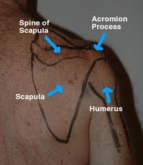
Traditional prolotherapy uses bony landmarks, which are palpated, marked, and then injected with an inflammatory agent. No imaging is used; hence, the ability to palpate bony structures and identify the underlying anatomy is important.
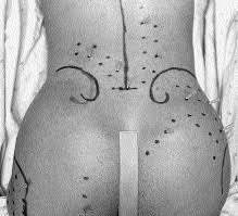
Since X-ray and ultrasound are NOT used, the areas to be injected are typically marked on the patient as shown below. Each dot represents a place where a needle is inserted without guidance and an inflammatory agent injected. Multiple small blind injections are performed in a grid-type pattern. Ouch!!
Because X-ray is NOT used and no contrast dye is injected, prolotherapy injections are not precise. A prolotherapy injection cannot confirm accurate placement of the needle into a specific joint and, therefore, cannot be considered diagnostic.
What Is the Newest Twist?
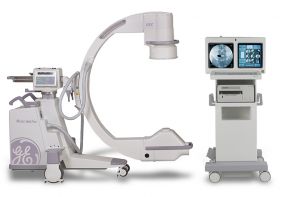
Many physicians who perform prolotherapy have recently purchased X-ray (fluoroscopy) units, which are used during the procedure. Unfortunately, they do not use contrast to confirm needle placement. Hence, these injections while performed under X-ray cannot be considered diagnostic.
What Does All This Mean?
Precision is required in the diagnosis and treatment of orthopedic conditions. In the cervical spine, for example, an injury to the C0/1 joint may be suspected based on history and clinical findings. The physician must accurately place a needle into the joint and confirm its placement with contrast that fills the targeted joint which is displayed below. This is the gold standard.

If you or a family member suffers from a cervical or lumbar spine condition or a common orthopedic condition, remember the gold standard as it relates to injections. An X-ray image of a needle in a joint that is filled with contrast is essential to be considered diagnostic and of clinical value. Anything less may delay clinical improvement and add additional time and expense to the treatment plan. At the Centeno-Schultz Clinic, all X-ray–guided injections are performed with contrast, which is the standard of care.