Ehlers-Danlos Syndrome (EDS)
In high school and college, your hyperflexibility was remarkable. The splits and advanced yoga postures were executed without difficulty or significant stretching. Then pain and joint instability became an issue limiting your activity. Your orthopedic specialist told you everything is fine and you simply suffered a sprain. Your PT thinks your may have a problem with your connective tissue. What is Ehlers-Danlos Syndrome? What are the common causes of Ehlers-Danlos Syndrome? Are there different types of Ehlers-Danlos Syndrome? What are the common symptoms? How is Ehlers-Danlos Syndrome diagnosed? What is the prevalence? What are the common treatment options for Ehlers-Danlos Syndrome? Are there new advanced treatment options? What areas can be treated with Regenerative injections? Is managing joint pain and instability caused by EDS possible? Let’s dig in.
Ehlers-Danlos Syndrome
Ehlers-Danlos Syndrome is a group of disorders that affect and weaken your connective tissue. Connective tissue exists in every part of your body. It provides support and structure to tissues and organs including bone, ligaments, tendons, blood vessels, and lymphatic vessels.
What Is Ehlers-Danlos Syndrome? (Connective Tissue Disease)
Ehlers Danlos Syndrome (EDS) is a group of inherited disorders that affect and weaken the connective tissues such as tendons and ligaments (1). It is a hereditary disorder which means you are born with it. EDS has many different signs and symptoms which can vary significantly depending upon the type of EDS and its severity. It most commonly affects the skin, joints, and blood vessels. Joints are typically hypermobile with excessive joint range of motion because of a defect in collagen formation.
Common Causes of Ehlers-Danlos Syndrome? (Inherited)
In most cases Ehlers-Danlos syndrome is inherited. That is to say that you are born with it. The two main ways EDS is inherited are:
Autosomal dominant inheritance:
The faulty gene that causes EDS is passed on by 1 parent.
Autosomal recessive inheritance:
The faulty gene is inherited from both parents.
All the genes listed below provide instruction on how to assemble collagen. Collagen is an important protein that serves as a major building block for bones, skin, muscles, tendons and ligaments. In patients with Ehlers-Danlos Syndrome there is defect in how the body makes collagen. The most common genes that cause EDS include:
- COL1A1
- COL1A2
- COL3A1
- COL5A1
- COL6A2
- PLOD1
- TNXB
Are There Different Types of Ehlers-Danlos Syndrome?
Yes! There are 13 major types of Ehlers-Danlos syndromes (2). Each type has a set of clinical criteria to help establish a diagnosis. Each type of Ehlers-Danlos Syndrome affects different areas of the body. All types of EDS however have one thing in common: hypermobility. Often there is significant symptom overlap between the EDS subtypes and other connective tissue disorders.
The three most common types of EDS are:
Hypermobile
Hypermobile EDS ( hEDS) is the most common form of EDS.
Classic
Classic is the second most common type of EDS. Previously it was also called EDS Type I & II.
Vascular
Vascular EDS is quite rare and is the most severe type of EDS. Vascular EDS is much different from Hypermobile and Classic EDS. In addition to loose joints, and translucent skin these patients are at risk for life-threatening rupture of the intestine, uterus, and arteries
The other types of EDS syndromes are:
- Cardiac-valvular EDS
- Kyphoscoliosis EDS
- Arthrochalasia EDS
- Dermatosparaxis EDS
- Musculocontractural EDS
- Myopathic EDS
- Periodontal EDS
- Brittle Cornea Syndrome (BCS)
- Spondylodysplastic E
Ehlers-Danlos Syndrome Symptoms And Clinical Presentations
EDS symptoms vary depending upon the specific type and its severity. There is significant clinical overlap between EDS types. The following is a general overview of the three most common EDS types and symptoms.
Hypermobile Ehlers-Danlos Syndrome
Patients with Hypermobile EDS may have:
- Joint hypermobility
- Unstable or loose joints that easily dislocate
- Fatigue
- Skin that is easy to bruise
- Digestive problems that include heartburn and constipation
- Problems with bladder control
Classical Ehlers-Danlos Syndrome
Patients with Classical EDS may have:
- Joint hypermobility
- Unstable or loose joint that easily dislocate
- Stretchy skin
- Fragile skin prone to injury
- Smooth, velvety skin that bruises easily
- Wounds that are slow to heal and leave wide scars.
Vascular Ehlers-Danlos Syndrome
Patients with this rare type of EDS can have involvement of the blood vessels and internal organs. They are susceptible to life threatening bleeding. Other symptoms include:
- Skin that is easy to bruise
- Thin skin with visible small blood vessels
- Fragile blood vessels that are prone to damage with bleeding
- Joint hypermobility
- Organs at risk for tearing (uterus and lung)
Associated Symptoms
Ankle Pain After Walking
More than any other joint, our ankles bear the burden of all of our body weight. The ankles aren’t large weight-bearing joints, like our hips or knees; comparatively speaking, the ankles are rather small for the tasks they are assigned to do. If the ankles are normal and healthy and there isn’t a weight issue placing excess stress on the ankles, the ankles can typically bear the forces of walking, running, hiking, and so on quite well. However, when the ankles are weak or carrying too much weight, any additional forces placed upon them—even something as simple as walking—can create problems. Do your ankles get sore after walking? What about foot and ankle pain after hiking? If so, it’s a good idea to proactively address it now, before it gets worse, rather than resigning yourself to it and decreasing or stopping the activities you enjoy. We’ll explain more in a moment, but first let’s take a closer look at the structure of the ankle.
Read More About Ankle Pain After WalkingAnkle Pain When Running
There are multiple causes of ankle pain while running. The six major causes are: Stress Fracture A stress fracture is a small crack in the bone due to overuse and repeated impact. They are a common cause of pain in runners, accounting for up to 16% of injuries. The shin bone (tibia) is the most commonly affected bone accounting for approximately 40 % of stress fractures. Pain is the most common symptom. Plantar Fasciitis. The plantar fascia is the thick connective tissue that extends from your heel to your toes. Plantar fasciitis is the inflammation of the plantar fascia and is the most…
Read More About Ankle Pain When RunningBack of Knee Swollen
Back of knee swollen? Swelling in the back of the knee is not something that is talked about too often. It can be uncomfortable or sometimes painful with the movement of the knee or, even with rest. It often stems from an orthopedic-related issue or, perhaps some other medical condition. Let’s dive in…What’s Causes Swelling in the Back of Your Knee? The back of the knee contains a variety of anatomical structures that can be affected and result in pain, swelling, numbness, tingling, etc. Muscles and tendons behind the knee that could be the source of pain or pathology could be one or more of the following: Gastrocnemius, Soleus…
Read More About Back of Knee SwollenBig Toe Numbness
Believe it or not, one of those significant issues that can present as numbness stems from the low back. In the lumbar spine, the nerve that exits the spine at the L5 level branches down through the hip, thigh, knee, lower leg, and, yes, all the way into the foot and toes. So a pinched or irritated nerve at that L5 level in the back can create problems, such as pain, numbness, tingling, and so on, anywhere along the nerve branch. So what can irritate the L5 spinal nerve? The list is long, but it includes disc issues, such as herniated…
Read More About Big Toe NumbnessBrain Fog
Brain fog is one of the hallmark symptoms of Craniocervical instability. It is characterized by slow thinking, difficulty focusing, confusion, lack of concentration, forgetfulness, or haziness in thought. The severity of symptoms varies from patient to patient. Ever had a bad hangover or high fever and had trouble concentrating or completing simple tasks? This brain fog. Some patients describe it as a generalized haziness in thought as if cotton were stuck in the head. In addition to cognitive impairments, many patients also report generalized mental fatigue making complex tasks almost impossible. The exact cause of brain fog is unknown. Possible explanations include…
Read More About Brain FogButt Pain
Throwing this question out to the social-media masses is sure to bring out the cyber “comedians” among us, but the truth is when you really do have butt pain, it’s no laughing matter. So we’re glad you came here to ask because we actually do see many patients in our clinic with this problem. There are a number of issues that can cause butt pain, but the root causes we most often discover are hamstrings tendinopathy, a pinched low-back nerve, or sacroiliac joint syndrome. Let’s take a look at each of these individually. – Hamstrings Tendinopathy: The hamstrings are actually a collection of large muscles…
Read More About Butt PainCervicalgia / Neck Pain
Cervicalgia is also known as neck pain, which is an all-too-common, unpleasant pain. Read here to learn the symptoms, diagnosis, and treatment options. We will discuss other causes for cervicalgia. What symptoms are associated with cervicalgia? How is cervicalgia diagnosed? What are the treatment options for cervicalgia? Cervicalgia is a medical term used to describe neck pain. It is very common and affects approximately 2/3 of the population at some point in their life. Cervicalgia is the 4th major cause of disability. Risk factors include injury, prior history of neck and musculoskeletal pain, jobs that require a lot of desk work, low social support, job insecurity, physical weakness, and poor computer station setup.
Read More About Cervicalgia / Neck PainChronic Fatigue
Have you ever had a challenging all-day hike, athletic endeavor, or worked a double shift and felt exhausted the next day? Later that day or the next morning, moving across the room may have seemed almost impossible and took herculean power. Now image this occurs on a daily basis without any provocation. 24/7 simple tasks are nearly impossible due to a lack of strength and energy. This is chronic fatigue, and it is one of the common symptoms associated with craniocervical instability. Severity can vary and in severe cases, patients are confined to their beds. Aggravating and alleviating factors often times can not be identified.
Read More About Chronic FatigueChronic Headaches
Do you suffer from chronic headaches? Maybe it’s a daily issue, maybe once a week, maybe even less often, but one thing’s for sure—when a chronic headache kicks in, it can be a real pain in the neck, literally. In order to effectively address chronic headaches, you have to first determine if the pain is caused by a problem in your neck. Let’s take a look at a few neck issues that can cause headaches: Weak neck muscles – The head, on average, weighs about ten pounds, so when the neck muscles are weak, it can make your head feel a bit like a bowling ball that your neck can’t quite balance. There are many muscles that, along with the cervical spine, work together to help support the neck and aid movement…
Read More About Chronic HeadachesEDS Back Pain
Low back pain is common throughout the United States with an incidence of 1.39 per 1000 person-years. It accounts for 3.15 of all emergency visits. It is also problematic in patients with Ehler-Danlos Syndrome (EDS). EDS back pain is truly a debilitating issue that could use some light shed upon it. A group of inherited disorders that affect and weaken the connective tissues such as tendons and ligaments (2). Primary sites of involvement include skin, joint, and blood vessels. Joints are typically hypermobile with excessive joint range of motion as a result of a defect in collagen formation. Overall, approximately 90% or more EDS patients suffer from chronic pain.
Read More About EDS Back PainEDS Headaches
Due to an inherent disorder in the brain blood flow patients with EDS are prone to migraines. Ehler-Danlos syndromes are classified into thirteen subtypes. Migraines with or without aura are the most common form of headaches in The EDS, hypermobility type (EDS-HT). A recent study demonstrated that in the EDS-HT group, migraines had an earlier onset and a higher number of episodes per month when compared to non-EDS patients. EDS patients commonly suffer a variety of headache types which include: tension headache, Occipital neuralgia due to pressure and irritation of the lesser and greater occipital nerves, Facet headache pain due to the instability of the facet capsule and ligaments ligament.
Read More About EDS HeadachesEDS Shoulder Pain
Shoulder pain can arise from many injuries including dislocation and subluxation. For patients with Ehler-Danlos Syndrome (EDS), this is especially problematic given their joint laxity. EDS is a group of inherited disorders that affect and weaken connective tissues such as tendons and ligaments (1). And EDS shoulder pain is often characterized by joints that are typically hypermobile with an excessive range of motion. Hypermobility means that the joint is too mobile or too lax, and it is a major culprit in shoulder pain. Ligaments and muscles stabilize joints and keep them moving in set predetermined motions with micro-millimeter precision. When the ligaments are loose. the joint surfaces crash together…
Read More About EDS Shoulder PainFibular Head Pain
The proximal fibula is the part of the bone that lives just below the knee joint on the outside. It’s attached to the leg bone (tibia) via strong ligaments and there is a small joint here. There are many things that attach here, so it’s a critical point where pain can occur. There is a small joint between the fibula and the tibia known as the proximal tibiofibular joint. This is a plane-type joint which allows some sliding of the fibula on the tibia. It has cartilage just like the knee joint, so it can get arthritis which means worn down cartilage and bone spurs.
Read More About Fibular Head PainFront of Hip Pain
Arthritis might be your first thought when you’re experiencing chronic pain at the front of your hip, but the truth is, there are many different issues that could be causing that hip pain. The ultimate goal, of course, is to fix the pain, but eliminating the pain once and for all involves also fixing the problem that is causing it…not just applying a temporary solution for pain relief. 5 Possible Causes for Front of Hip Pain Today we’re going to explore the hip and 5 possible causes for pain specifically in the front of the hip as well as cover ways you can test for each.
Read More About Front of Hip PainGluteus Medius Pain
The gluteus medius is one of the major muscles in your buttock and hip. There are three gluteal muscles you have probably heard about: They layer one upon the another much like a layer cake. The gluteus minimus is closest to the body followed by the gluteus medius and then the larger gluteus maximus.Gluteus Minimus – The gluteus minimus is the smallest of the three gluteal muscles. It is a small triangular muscle that lies underneath its bigger brother, the gluteus medius. It functions to stabilize the hip, rotate the thigh, and move the hip in an outward direction.Gluteus Medius -A powerful muscle that starts at the backside of your waist bone…
Read More About Gluteus Medius PainIlium Pain
There are many different causes of ilium pain. For ease, I have broken them down into two groups: Direct and Referred. Direct causes are those directly related to the ilium bone itself and the structures that attach to it. Referred pain is pain that is felt or perceived in a part of the body other than its actual source. The classic example of referred pain is someone who is having a heart attack and feels pain radiating down their arm. The pain is felt or perceived in the arm while the real tissue injury is occurring in the heart. Direct causes of ilium pain include…
Read More About Ilium PainKnee Buckling
Knees can buckle, causing a sensation of one or both knees giving out that affects nearly 17% of adults. The knees are regarded to be one of the most essential (and biggest) joints in the human body because they play significant roles in basic activities such as walking and sitting. When our knees begin to feel unstable, weak, or begin giving out, it is easy to become stressed since we rely so heavily on them for movement. The knee can become stable from numerous types of injuries. The knee is kept stable by the following structures: Tendons attach the leg muscles…
Read More About Knee BucklingKnee Clicking
Clicking in the knee can occur for a number of reasons. The 5 major causes include: ligament injuries, meniscus tears, knee arthritis, patellofemoral syndrome, and soft/scar tissue. Let us start with ligament tears. A ligament is a thick piece of connective tissue that connects one bone to another. The knee has 4 principal ligaments: ACL, PCL, MCL, and LCL. Ligaments provide important stability for the knee (1). If injured or torn, ligaments can create instability. This instability can cause clicking in the knee. As ligaments travel across boney surfaces they can also catch make the knee click. The meniscus is a C-shaped cushion situated between the thigh bone and shin bone.
Read More About Knee ClickingKnee Hurts When I Bend It and Straighten It
Your knees bend countless times throughout the day. Running up the stairs, down the hall after kids, and getting into the car. You straighten the knee as you walk, descend stairs or get into and out of the car. Bending and straightening the knee are necessary for daily activities. Knee pain with bending or straightening may be a mild, transient irritation or may indicate a more significant problem. Learn more below and avoid further injury and dysfunction. Knee pain can vary significantly depending upon many factors including the actual source of the pain, the severity of the injury, general health, and level of activity…
Read More About Knee Hurts When I Bend It and Straighten ItKnee Hyperextension
The knee is a hinge joint between the femur and the tibia along with an accessory bone, the fibula. These bones are held together via ligaments that keep the bones aligned while the joint goes through its natural range of motion. These ligaments are: Medial Collateral Ligament (MCL), Lateral Collateral Ligament (LCL), Anterior Cruciate Ligament (ACL), Posterior Cruciate Ligament (PCL). from zero degrees fully extended to 140 degrees fully flexed. BUT when a force from the front to the back overcomes the strength of the ligaments the knee is forced into an unnatural position called “Hyperextension” which, in turn, can…
Read More About Knee HyperextensionKnee Locking & Catching
Knees have the ability to bend forward and back, as well as rotate slightly. When a knee is unable to execute these actions, it impairs mobility and the capacity to complete daily chores such as sitting, standing, squatting, or kneeling. A locked knee occurs when a knee cannot be bent or straightened so it gets stuck or feels locked in a certain position for an extended period of time. There are two forms of locked knees: one that is due to inability to move because of an actually physical or mechanical restriction and there is one that feels locked due to the pain involved in moving it. When a person has their knee joint effectively frozen into place and cannot move, this is known as a true locked knee….
Read More About Knee Locking & CatchingLeg Feels Disconnected
So what exactly causes the leg to feel disconnected? The hip joint is the connection point between the leg and the rest of the body. The femur in the leg and the acetabulum in the pelvis create the hip joint. So if the left SI joint, for example, in the back and the symphysis pubis joint in the front becomes unstable due to loose supporting ligaments, then the left leg, which is connected between the two joints, is likely to also feel a bit disconnected. Likewise, if the right SI joint were unstable, then the right leg might feel disconnected. How This Patient’s “Disconnected” Leg Was Fixed Despite “leg feels…
Read More About Leg Feels DisconnectedLoose Knee Joint
Ligaments are fibrous bands that connect the bones together. If the ligaments inside your knee become damaged, your knee could feel loose (with or without pain). While walking, exercising, or simply standing, it may feel like your knee could collapse at any moment. If the knee muscles are damaged, they will be unable to contract properly, resulting in lack of strength and discomfort. Swelling might have a similar result. What Causes Laxity in the Knee Joint? Knee injuries are generally sports-related, although they may occur during any physical activity that puts stress on the knee and causes bending or trauma…
Read More About Loose Knee JointLower Back Pain When Sitting
After a long day on your feet sitting down is supposed to be way to relaxing. Unfortunately for some sitting for any length of time can be painful. Most people experience low back pain at some point in their life. The lifetime prevalence of low back pain is 85% (1). Let’s take a deeper look at the different types of pain and causes of low back pain when sitting. Pain can present in many different ways. It can be intermitent or constant. The quality of the low back pain can also vary depending upon the actual source of injury. Common examples include: Sharp and Stabbing, Dull and Aching, Throbbing/ Pulsating, Pins and Needles, Burning, Electrical
Read More About Lower Back Pain When SittingLower Back Pain When Standing
When you’re seated, the facet joints in your lower back are in an open and slightly flexed position. When you stand up, these joints compress. If they are painful or have arthritis, you’ll have pain as you stand up because this puts pressure on the painful joints. In addition, if there is any type of movement of one vertebra forward on another (called spondylolisthesis), then this shift will have occurred as you sit. This is called degenerative spondylolisthesis. When you get back up, the vertebrae will come back into position after a few seconds, leading to that awkward “walk it out period” that starts out painful and ends up more normal.
Read More About Lower Back Pain When StandingNeck Pain And Dizziness
Our body’s balancing system is compromised of three separate systems that work closely together to keep the body in balance: the eyes, inner ear, and upper cervical spine. The upper cervical spine contains and processes information about your position in space and communicates this information to your eyes and inner ears via nerves. There is a constant highway of electrical signals between the cervical spine, inner ear, and eyes that keep us upright, make us aware of our position and enable us to walk, move and run. This balancing system requires the inner ear, eyes and cervical spine to be operational…
Read More About Neck Pain And DizzinessOuter Hip Pain
The hip is a ball-socket joint. The ball is the top part of the thigh bone (femur). The socket is formed by the ilium bone which is one of three bones that make up the pelvis. The ball-socket joint moves in different directions and is lined with cartilage. The actual hip joint is located approximately at the crease between your stomach and top of your thigh. This area is also known as the groin. The top of the ilium bone is called the iliac crest (waist bone). If you firmly place your hand on your waist you can feel a bone that spans your side.
Read More About Outer Hip PainOutside Ankle Pain
We often see patients with outside ankle pain who have no idea how it happened; they just know their ankle suddenly started hurting. One patient was a classic example of this. As an athletic hockey and Lacrosse player, however, it’s highly possible he experienced sprains and other ankle injuries, even minor ones, over the years, and these, rather than one big traumatic episode, could have been the catalyst to his sudden outside ankle pain. Outside ankle pain can be treated without surgery by an interventional orthopedics physician. Loose or torn ligaments usually can be treated nonsurgically with ultrasound-guided high-dose platelet-rich plasma (PRP) injections…
Read More About Outside Ankle PainOutside Knee Pain
What causes outside knee pain? How do you treat it? Let us go over all of this and also review how one patient avoided a huge surgery. What is On the Outside of the Knee? You have a couple of key structures here (1). They include the: Lateral meniscus, Lateral joint compartment, Popliteus tendon, Iliotibial band, Lateral collateral ligament , and anterolateral ligament, and Fibula. The meniscus is a figure-8 shaped fibrous structure (shown here from above) that is a shock absorber for the knee joint. It has an outside part (lateral meniscus) that cushions the joint. The meniscus can become torn or degenerated. If you are young…
Read More About Outside Knee PainShoulder Cracking
The Scapula is your shoulder blade. It is a large triangular bone that compromises part of the shoulder joint. The Scapula is positioned over the back chest wall and ribs where it moves in different directions with shoulder movements (1). The Scapula meets with the arm bone (humerus) to form the shoulder joint. It also meets with the clavicle to form the AC joint. Multiple structures attach onto the Scapula which provides important support and movement for the scapulae and shoulder joint (2). These include:- Muscles/Tendons: Multiple muscles attach to the Scapula which include the deltoid, supraspinatus, infraspinatus, triceps, and teres minor.
Read More About Shoulder CrackingShoulder Pain When Reaching Across Body
Shoulder pain can make simple chores almost impossible. Have you ever reached for an object high on a shelf only to have pain that takes your breath away? What causes shoulder pain when reaching across the body? What is shoulder impingement? What does shoulder impingement feel like? Can a shoulder X-ray show shoulder impingement? What are the treatment options for shoulder pain when reaching across the body? ulder impingement and rotator cuff injuries are among the most common causes of shoulder pain (1). Both can cause shoulder pain when reaching across the body. Shoulder impingement is a painful condition in which the bursa and muscles of the shoulder are pinched or compressed.
Read More About Shoulder Pain When Reaching Across BodyShoulder Pain When Sleeping
There are several reasons why shoulder pain at night occurs or is aggravated; The common explanations include: Sleep typically involves a static position lasting hours at a time with little or no movement. Sleeping on your side places additional pressure on the tendons and bursa of the shoulder. Sleep can cause muscles and tendons to settle in a slightly different position resulting in additional pressure and reduced blood flow. Identifying the underlying problem is important! This allows therapy to focus exclusively on the exact problem or problems. When appropriate conservative care is always the first line of treatment. Focus is typically on strengthening and improving range of motion.
Read More About Shoulder Pain When SleepingShoulder Popping
Noises in the joints, such as popping, cracking, or clicking, can be quite disturbing and cause concern. That popping feeling is called crepitus. That’s something to be ignored, right? It certainly seems harmless enough—after all, there’s no pain, just that annoying popping when you move your shoulder this way or that. But just because there’s no pain doesn’t mean there’s no problem. There’s actually good news and bad news here. The good news? All that shoulder popping really could be no big deal. The bad news? That popping could be slowly chewing up your shoulder joint, and it may be a warning sign that arthritis is on its way.
Read More About Shoulder PoppingSI Joint Pain
The SI Joint is an important joint in the lower back that can cause pain and dysfunction. Pain is typically one-sided involving the low back and buttock but when severe can radiate down the leg. Each of us has a right and left SI joint. The pelvic ring consists of the two SI Joints and the symphysis pubis. Each is rich in supporting ligaments that provide stability for the joint. SI Joint pain is aggravated by ligament instability and muscle weakness. Physical therapy is the first line of treatment for SI Joint injuries. For best clinical results the stability of the joint and muscle strength must be evaluated and treated…
Read More About SI Joint PainStiff Neck
Conventional wisdom states that stiff neck symptoms can be treatable with nonsurgical remedies. However, not all stiff neck symptoms are benign, and leaving the stiff neck untreated can lead to a limited range of motion that can affect your overall health and quality of life. A stiff neck can cause pain, tightness, popping, and clicking noises and sensations and affect daily activities. A detailed examination of stiff neck symptoms can determine the exact condition affecting the stiffness of one’s neck. Where does the neck hurt? Neck stiffness can occur at the base of the head, down to the shoulders. This can be associated with pain, crunching noises, or popping sensations and may…
Read More About Stiff NeckTight Hamstrings & Calves
The hamstrings are a group of muscles in the upper leg. They are located on the backside of the upper leg and are comprised of three muscles: biceps femoris, semitendinosus, and semimembranosus. The calf muscles are located on the backside of the lower leg and are comprised of two muscles: the gastrocnemius and soleus. Tight hamstring and calf muscles can be painful, limiting someone mobility. They also make lower extremity muscles more vulnerable to injuries. Tight hamstrings and calves can arise from different sources which include: medication, muscle and tendon injury, overuse, muscle imbalance, dehydration, poor posture, low back injury…
Read More About Tight Hamstrings & CalvesDiagnosing EDS
Establishing a diagnosis of Ehlers-Danlos Syndrome starts with a full history and physical examination. While your PCP may start the process, a referral to Rheumatologist or another specialist is common. During the physical examination both the skin and joints are examined in detail. How far the skin is able to be stretched ( hyperextensibility) is important as is the flexibility of the joints. A Beighton score is often times utilized. It is a simple system of maneuvers that quantifies joint laxity and hypermobility (3). It uses a 9-point system where the higher the score the higher the laxity. Other testing may include:
- Collagen Typing
- Genetic Testing
- MRI
- CT (Computerized Tomography)
- Ultrasound of the Heart (Echocardiogram)
- Urine Analysis
- Skin Biopsy
The Prevalence of EDS
The estimated prevalence for all EDS varies between 1/10,000 and 1/25,000 (5). Hypermobile EDS is more common than previously reported (6)
Common EDS Treatment Options
Specific treatment will depend upon the specific type of the EDS and its severity. EDS is not curable but certainly treatable. When appropriate conservative therapy should always be the first option. Treatment options include (4):
Physical Therapy
PT can provide specific exercises and workouts that focus on joint strength. The stability of a given joint is dependent upon muscle strength and ligament integrity. In patients with EDS muscular strengthening can enhance joint stability thereby limiting dislocations, injuries and pain.
Bracing
External bracing is oftentimes utilized in an effort to provide stability when muscle strength and ligamental stability are compromised. The goal is to stabilize the joint so as to prevent injuries, pain, and dysfunction. Examples include knee and ankle braces. In more advanced cases wheelchairs and scooters may be necessary.
Surgical Procedures
Surgery may be necessary when life-threatening conditions occur such as organ rupture, uncontrolled bleeding and retinal detachment occurs.
Regenexx Procedures
Regenexx is a highly specialized, research-based group of nonsurgical procedures that use PRP and Bone Marrow Concentrate to treat common orthopedic conditions. Bone Marrow Concentrate contains stem cells. This will be discussed in detail below.
There’s A Non-Invasive Way To Manage Pain
There are several ways to manage the pain depending upon the source and its severity. When conservative care in the form of PT, OT and Chiropractic care fail, there are other available options which include
Medications
Good quality fish oil and Tumeric can address the pain and inflammation. NSAIDs should be avoided due to associated risks and negative impact on cartilage and stem cell function. Oral steroids should also be avoided as they can adversely affect ligament stability.
Injections
Not all injections are the same. Steroid injections should be avoided in patients with EDS as they can weaken the already compromised ligaments. They can make the joint hypermobility and dysfunction worse.
At the Centeno-Schultz Clinic, we are experts in the treatment of ligament injuries. Treatment options include the following:
Prolotherapy
Prolotherapy, also known as proliferation therapy, involves the injection of an irritant such as dextrose into a damaged or weakened structure. These are typically ligaments. The injected irritant initiates the body’s innate healing process by increasing inflammation. The increased inflammation triggers the body to send healing cells, platelets, and specialized proteins to the area. The intended result is the body’s healing and strengthening of the injected structures.
PRP
PRP stands for Platelet Rich Plasma. Platelets are cells within our blood that are responsible for clotting. They are also rich in growth factors that can increase blood flow in a given area thereby promoting healing. PRP can be made in a lab or by a bedside automated centrifuge. Blood is drawn from a patient’s arm and processed in the lab or automated centrifuge. Certain fractions of the blood are removed allowing the platelets to be super concentrated. At the Centeno-Schultz Clinic, we have a state-of-the-art laboratory that allows us to customize the PRP dose to a specific patient’s need. Think of growth factors like espresso shots for local repair cells. They stimulate other cells to do more work and can even call the needed repair cells to the area.
Bone Marrow Concentrate
Bone Marrow Concentrate contains your own stem cells. It is an Autologous procedure because the bone marrow comes from you. In contrast Allogenic means bone marrow from a person other than the patient. Bone marrow is commonly taken from your iliac crest (hip) using a needle to remove the bone marrow. The procedure is performed under x-ray or ultrasound guidance. A local anesthetic is injected into the skin and deeper structures prior to the aspiration. The bone marrow aspirate is then placed into a centrifuge where the cells are separated.
What Areas Can be Treated with Regenerative Injections?
At the Centeno-Schultz Clinic we acknowledge the complexity of the human body and recognize that for best clinical outcomes it is best to view and treat the body as a whole. This concept is referred to as Functional Spinal Unit (FSU). To learn more about this approach please click here.
The list of areas that can treated in patients with EDS is extensive. The most common include:
Cervical, Thoracic and Lumbar Spine
Supraspinous, Interspinous and Thoracolumbar fascia
Upper Cervical Ligaments
Alar and Transverse ligaments
Sacroiliac Joints and Ligaments
Knee Ligaments
Knees and important ligaments that include ACL, PCL, MCL, and LCL
Hip Ligaments
Hips and supporting capsule and ligaments
Shoulders
Superior, middle and inferior glenohumeral ligaments
Ankles
Deltoid, Anterior Talofibular and Calcanofibular ligaments.
The doctors at Centeno-Schultz Clinic are experts in the treatment of patients with EDS and hypermobility. We have pioneered the following procedures to address important ligament injuries.
Knees
Nonsurgical treatment of Anterior Cruciate Ligament tears in the Knee utilizing both PRP and Bone Marrow Concentrate. To learn more please watch the video below.
Upper Cervical Ligaments
Important ligaments connect your head to your cervical spine. The two most important are the Alar and Transverse ligaments. Patients with Ehlers-Danlos Syndrome can injure or stretch these ligaments giving rise to a number of symptoms that oftentimes are poorly understood and recognized. The condition is called Craniocervical Instability and has been discussed in prior blogs. To learn more about this revolutionary nonsurgical treatment option please click on the video below.
Managing Pain Brought On By Ehlers-Danlos Syndrome Is Possible!
Ehlers-Danlos Syndrome can be associated with joint instability, pain, and dysfunction. When conservative care fails to provide significant clinical improvement, the good news is that there are new advanced treatment options. Injured, stretched, or partially torn ligaments and tendons can be precisely injected under MSK ultrasound with Prolotherapy, PRP, or Bone Marrow Concentrate. Injections without guidance also known as blind injections are below the standard of care at the Centeno-Schultz Clinic and often miss the targeted tissue and fail to provide patients with the best clinical outcomes.
In Conclusion
- Ehlers-Danlos Syndrome (EDS) is a group of inherited disorders that affect and weaken the connective tissue such as tendons and ligaments.
- Most cases of EDS are inherited.
- There are 13 major types of EDS.
- The three most common types of EDS are Hypermobile, Classic and Vascular.
- Symptoms and clinical presentations vary depending upon the specific type and severity.
- Diagnosing Ehlers-Danlos Syndrome involves history and physical examination. A specialist is often times used to confirm the diagnosis. Testing may include collagen testing, genetic testing, MRI, CT, Echocardiogram, urine analysis and skin biopsy.
- Common treatment options include physical therapy, bracing and medications. Life threatening conditions can warrant surgery.
- Precise injections of Prolotherapy, PRP or Bone Marrow Concentrate can potentially accelerate healing and enhance stability of hypermobile joints.
- Areas that can be treated include the cervical, thoracic, and lumbar spine, upper cervical ligaments, sacroiliac joints, knees, hips, shoulders and ankles.
If you or a loved one suffers from Ehlers-Danlos Syndrome and have persistent joint pain and limitations despite conservative care, please schedule a telephone candidacy discussion with a board-certified, fellowship-trained physician. We can help you live comfortably with your Ehlers-Danlos condition. Learn about our advanced regenerative procedures that can improve your quality of life and get you back in the game.
Our Doctors Who Treat Pain Associated with EDS

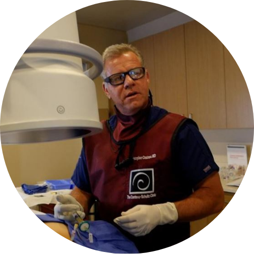
Christopher J. Centeno, MD
Christopher J. Centeno, M.D. is an international expert and specialist in Interventional Orthopedics and the clinical use of bone marrow concentrate in orthopedics.
Dr. Centeno is one of the few physicians in the world with extensive experience in the culture expansion of and clinical use of adult bone marrow concentrate to treat orthopedic injuries. His clinic incorporates a variety of revolutionary pain management techniques to bring its broad patient base relief and results. Dr. Centeno treats patients from all over the US who travel to Colorado to undergo innovative, non-surgical treatments. Dr. Centeno has chaired multiple international research-based conferences. He also maintains an active research-based practice, with multiple publications listed in the US National Library of Medicine. Dr. Centeno has also served as editor-in-chief of a medical research journal dedicated to traumatic injury.
Dr. Centeno trained at the Baylor College of Medicine, Texas Medical Center, and the Institute for Rehabilitation Research. He hails from both Florida and New York and currently resides in Boulder, Colorado with his wife and three children.

John Schultz, MD
John R. Schultz M.D. is a national expert and specialist in Interventional Orthopedics and the clinical use of bone marrow concentrate for orthopedic injuries. He is board certified in Anesthesiology and Pain Medicine and underwent fellowship training in both. Dr. Schultz has extensive experience with same day as well as culture expanded bone marrow concentrate and sees patients at the CSC Broomfield, Colorado Clinic, as well the Regenexx Clinic in Grand Cayman. Dr. Schultz emphasis is on the evaluation and treatment of thoracic and cervical disc, facet, nerve, and ligament injuries including the non-surgical treatment of Craniocervical instability (CCI). Dr. Schultz trained at George Washington School of…
Read more
John Pitts, M.D.
Dr. Pitts is originally from Chicago, IL but is a medical graduate of Vanderbilt School of Medicine in Nashville, TN. After Vanderbilt, he completed a residency in Physical Medicine and Rehabilitation (PM&R) at Emory University in Atlanta, GA. The focus of PM&R is the restoration of function and quality of life. In residency, he gained much experience in musculoskeletal medicine, rehabilitation, spine, and sports medicine along with some regenerative medicine. He also gained significant experience in fluoroscopically guided spinal procedures and peripheral injections. However, Dr. Pitts wanted to broaden his skills and treatment options beyond the current typical standards of care.
Read more
Jason Markle, D.O.
Post-residency, Dr. Markle was selected to the Interventional Orthopedic Fellowship program at the Centeno-Schultz Clinic. During his fellowship, he gained significant experience in the new field of Interventional Orthopedics and regenerative medicine, honing his skills in advanced injection techniques into the spine and joints treating patients with autologous, bone marrow concentrate and platelet solutions. Dr. Markle then accepted a full-time attending physician position at the Centeno-Schultz Clinic, where he both treats patients and trains Interventional Orthopedics fellows. Dr. Markle is an active member of the Interventional Orthopedic Foundation and serves as a course instructor, where he trains physicians from around the world.
Read more
Brandon T. Money, D.O., M.S.
Dr. Money is an Indiana native who now proudly calls Colorado home. He attended medical school at Kansas City University and then returned to Indiana to complete a Physical Medicine and Rehabilitation residency program at Indiana University, where he was trained on non-surgical methods to improve health and function as well as rehabilitative care following trauma, stroke, spinal cord injury, brain injury, etc. Dr. Money has been following the ideology behind Centeno-Schultz Clinic and Regenexx since he was in medical school, as he believed there had to be a better way to care for patients than the status quo. The human body has incredible healing capabilities…
Read moreOther Resources for EDS
-
Craniocervical Instability & Ehlers Danlos Syndrome: Know the Facts
Gymnastics and yoga were easy as you were super flexible. With continued training your shoulders and knees kept popping out of place. A series of small but significant traumas left you sidelined both from athletics, academics and socially. Brain fog and fatigue were common themes. You have seen countless physicians with conflicting information. Your chiropractor…
-
CSF Flow Measurement And Recovery From CCJ Instability
We’re nearing the quarter-century point of the 21st century, and technology in the field of medicine continues to advance at an incredibly fast pace. Thanks to advances in imaging technology, we’ll learn more in the following few decades about the neuromusculoskeletal system than we’ve learned in the past few hundred years. A clear example of…
-
The Amazing Collapsing Boy
Our children are collapsing under the weight of gravity. Why? They look down all day at screens, and it just wreaks havoc on our posture. For some, this is a major problem now, and for others, this will be a problem in the future. So this morning I’ll talk about Seth, whom I’ll call the…
———————————
1.Chopra P, Tinkle B, Hamonet C, et al. Pain management in the Ehlers-Danlos syndromes. Am J Med Genet C Semin Med Genet. 2017;175(1):212-9.DOI: 10.1002/ajmg.c.31554
2.Malfait F, Castori M, Francomano CA, Giunta C, Kosho T, Byers PH. The Ehlers-Danlos syndromes. Nat Rev Dis Primers. 2020 Jul 30;6(1):64. doi: 10.1038/s41572-020-0194-9. PMID: 32732924.
3.Smits-Engelsman B, Klerks M, Kirby A. Beighton score: a valid measure for generalized hypermobility in children. J Pediatr. 2011 Jan;158(1):119-23, 123.e1-4. doi: 10.1016/j.jpeds.2010.07.021. Epub 2010 Sep 17. PMID: 20850761.
4.Song B, Yeh P, Nguyen D, Ikpeama U, Epstein M, Harrell J. Ehlers-Danlos Syndrome: An Analysis of the Current Treatment Options. Pain Physician. 2020 Jul;23(4):429-438. PMID: 32709178.
5.Germain DP. Ehlers-Danlos syndrome type IV. Orphanet J Rare Dis. 2007 Jul 19;2:32. doi: 10.1186/1750-1172-2-32. PMID: 17640391; PMCID: PMC1971255.
6.Demmler JC, Atkinson MD, Reinhold EJ, Choy E, Lyons RA, Brophy ST. Diagnosed prevalence of Ehlers-Danlos syndrome and hypermobility spectrum disorder in Wales, UK: a national electronic cohort study and case-control comparison. BMJ Open. 2019;9(11):e031365. Published 2019 Nov 4. doi:10.1136/bmjopen-2019-031365


