The neck pain was unrelenting and unresponsive to conservative care. Simple tasks and motions were impossible. At times there were sharp, shooting pains radiating down your arm with accompanying numbness and tingling. Your doctor ordered a Cervical MRI and you are awaiting the results. What is a Cervical MRI? How does an MRI work? Does MRI imaging use x-ray? What are the differenes between a normal vs abnormal Cervical Spine MRI? What are the advantages of MRI based diagnoses? What are the shortcomings of traditional MRI imaging? When should you get a Cervial Spine MRI? What does a normal Cervical Spine MRI look like? What are the most common abnormal findings? What are the consequences of misinterpreting a Cervical Spine MRI? What alternative exists? Lets dig in.
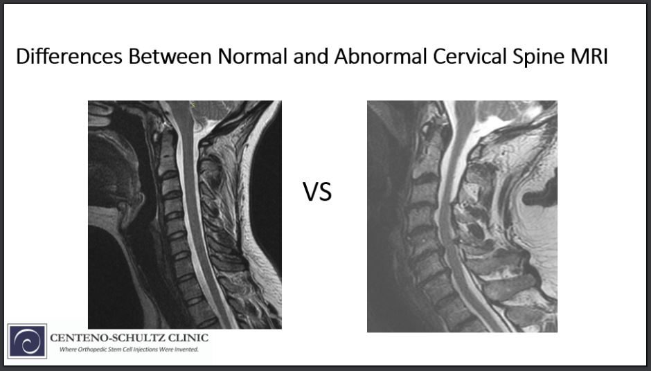
A Deeper Look into A Normal vs Abnormal Cervical Spine MRI
The internet has made medical information widely available. Many of our patients spend hours researching the results of their Cervical Spine MRIs. Self diagnosis is very common. Google is free whereas a medical education is many long years and hundreds of thousands of dollars. Reading your MRI report summary and researching the findings is not enough. There are important differences between a normal vs abnormal Cervical MRI which which will be discussed below.
What Is a Cervical Spine MRI?
MRI stands for Magnetic Resonance Imaging.
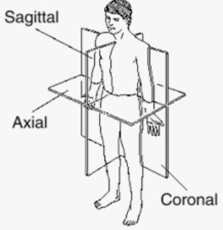
A Cervical MRI scan provides highly detailed pictures of your neck from different angles (1). The pictures are referred to as images and are numbered based upon the number of pictures taken. The different views include:
Axial: This is a cross section image. It divides the body into top and bottom halves. Image cutting the body in half with a top and bottom portion.
Coronal: This is a frontal view. it is a vertical plane that divides the body into front and back. I imagine a crown slicing downward creating a front and back.
Sagitial: This is a side view. Sagitial images start on the right side of the Cervical Spine and move to the left.
These different views allow physicians to accurately identify any and all abnormalities in your Cervical Spine MRI.
How Does an MRI Work?
MRI is an non-invasive technology that uses a powerful magnetic field and radio waves to produce detailed, 3-D pictures of your body (2). Our bodies contains millions of hydrogen atoms. Powerful magnets produce a strong magnetic field that forces the atoms in our body to align with that field. They all line up in the same direction. The radio waves the MRI produce disrupt this alignment. The radio waves are pulsed: turned on and then off. When they are turned off the MRI sensors are able to detect the energy released as the atoms realign with the magnetic field. This creates detailed images of our bodies.
An MRI scan uses a powerful magnetic field and radio waves to take detailed, 3-D pictures of your body.
Does a MRI Imaging Use x-ray?
No!
Advantages of MRI Based Diagnoses
MRI is a very powerful imaging modality that is being used with increased frequency. There are many advantages of being diagnosed with an MRI which include:
Detailed pictures of bones, cartilage, muscles, tendon and nerves. This is not possible with x-ray which only evaluates bone.
No radiation
Better resolution of soft tissues such as muscle, tendons and fat than CT scan
Detailed images from different views that allow better localization of a given structure. If you are evaluating a disc herniation for example you need to know exactly where it is, whether it is right or left sided and whether it irritates or compresses the exiting nerve root. An MRI can provide this specific information.
Shortcomings of Traditional MRI Based Diagnoses
Diagnosis Made in Isolation
Radiologists review MRI imaging and provide a formal report often times without any consideration of the patient’s medical history, limitations and current symptoms. Rarely are they aware of a patient’s entire medical history and findings on physical examination. Hence the diagnosis is made in isolation. Radiologists are obligated to report everything that they see. Hence the MRI report may mention different finding and abnormalities. Unfortunately all, some or none of the findings may explain a given patient’s symptoms.
Static vs Dynamic Studies
Traditional Cervical spine MRIs are performed with the patient lying still on their back. Movement in these type of MRI scans can compromise the quality of the images. These are known as static tests. Unfortunately these static tests can not evaluate the instability of the spine or the forces of gravity. Newer MRI technology now allows for patient to be upright as opposed to being flat. It also allows pateints to flex forward or extend backwards evaluating the presence or absence of instability. This is critical information that previously has not be available with static MRI imaging.
When Should You Get a Cervical Spine MRI? (Neck Pain)
There are many reasons why your doctor may order an MRI. The most common indication is severe neck pain or radiating arm pain that has not responded to physical therapy, rest, medications, chiropractic care.
Other reasons for a cervical spine MRI include
Trauma to the neck
Infection
Scoliosis (Curvature)
Headaches
Tingling and weakness in the arm
Birth defects of the spine
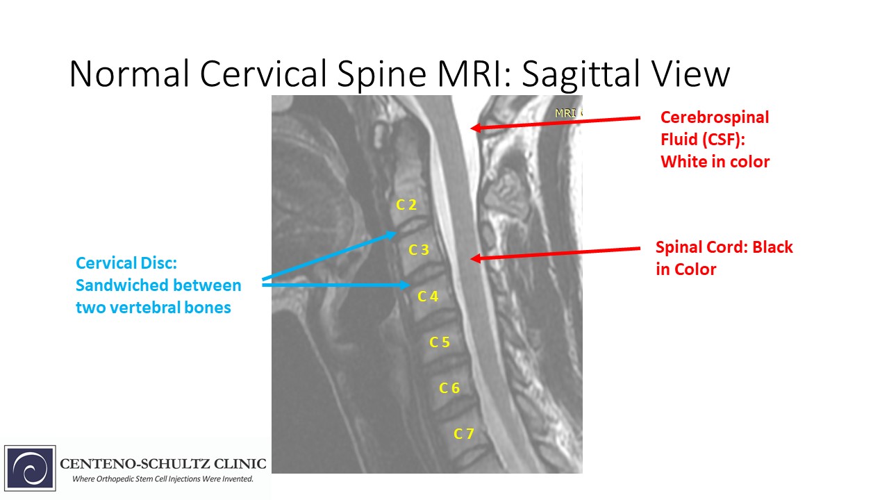
What Does a Normal Cervical MRI Look Like?
The Cervical Spine is composed of many different parts. Each is important and works in concert with all the other parts. The major parts of the Cervical Spine include:
Cervical Bones and Discs
The neck is composed of 7 boney building blocks that are called Vrtebral bodies. They are numbered from top to bottom starting with C1. The C denotes that we are discussing the cervical spine. Sandwiched between each of the bones is a a cushion also known as a Disc. The Disc is critical to the integrity and proper functioning of the neck as it absorbs the daily forces of living.
Cervical Facet Joints
A Facet Joint is a paired joint that resides in the back of the neck. They are cartilage lined joints that provide stability and limit range of motion. There is a right and left facet joint at each Cervical level. They are identified by the two vertebral bodies that they span. For example, the C5/6 facet joint is composed of the bones from both the C5 and C6 neck bones.
Central Canal
The Central Canal is a space that runs the entire length of the spine. It contains the spinal cord and spinal fluid. Narrowing of the Canal called stenosis and is graded based upon the severity of the narrowing. Central Canal Stenosis can cause several symptoms including neck pain, numbness of tingling in the arms and hands and problems with balance.
Neural Foramen
The Neural Foramen are the body’s doorways through which the spinal nerve roots exit the spinal column. There is a Neural Foramen at each level of the spine. There is also a right and left Neural Foramen. Narrowing of this boney doorway is called Neural Foramen Stenosis. This can cause irritation or pressure on the exiting nerve resulting in shooting pain down the arm, weakness and numbness.
Muscles
There is a large number of both large and small muscles in the neck. They play a critical role in the stability and mobility of the spine. Muscles can be injured, torn and atrophied (shrunken) and filled with adipose. This is especially important for the multifidus muscle which is a principal stabilizer in the spine. A previous blog discusses the importance of this small muscle in greater detail.
Tendons
Tendons are thick pieces of connective tissue that connect muscle to bone. Tendons are susceptible to inflammation, degeneration, partial thickness, and full thickness tears all of which can be visualized on a cervical spine MRI.
Common Abnormal Findings in Cervical Spine MRIs
The most common abnormal findings that you may come across in your Cervical Spine MRI report includes:
Cervical Spondylosis
This is a very broad term that is used to describe degenerative arthritis in the Cervical Spine. It is very common and typically gets worse with age.
Disc Injuries
The Disc is a gelatinous shock absorber that is sandwiched between each cervical vertebral bone. It absorbs the daily forces of life. It is susceptible to a number of different injuries which include Disc Protrusion, Herniation, and Extrusion.
Bone Spurs
A Bone Spur is a boney growth that develops on the edge of a bone. They can be small or large, smooth or irregular. They are typically the result of micro-instability. A Bone Spur is an attempt by your body to create stability. Bone Spurs in the cervical spine can compress nerves and narrow both the Neural Foramen and Central Canal creating Stenosis.
Facet Injuries
A Facet Joint is a paired joint that resides in the back of the neck. The joint is susceptible in injury and degeneration resulting in pain and restriction in range of motion. Trauma is the most common cause of injury.
Subluxation
The Cervical bones stack one upon another creating the Cervical Spine. The are all in alignment much like the drawers in your kitchen cabinets. Alignment allows for optimal performance and function. Unfortunately, trauma and wear and tear can compromise this alignment. Specifically, one vertebral bone can slip forward in relation to the other. This is called Anterolisthesis. “Listhesis” means slippage. Anterior means forward. A vertebral body can also slip backwards in relation to the other bones. This is called Retrolisthesis. Anterolisthesis and Retrolisthesis are both forms of Subluxation and can cause significant pain and dysfunction.
Central Canal Stenosis
The central canal is a space that runs the entire length of the spine. It contains the spinal cord and spinal fluid. Narrowing of the Canal space is called Stenosis and is graded depending on its severity. There are multiple causes of Central Canal Stenosis which include Disc Protrusions, Disc Herniations, Subluxations, Facet overgrowth and ligament thickening.
Foraminal Stenosis
Nerves exit the spinal cord through a boney doorway called the Neural Foramen. Foramen means opening or doorway. Stenosis means narrowing. Foraminal Stenosis is narrowing of the boney doorway through which the spinal nerves exit. When this occurs, the nerve can be irritated or compressed resulting is pain, numbness, tingling and weakness.
Consequences of Misinterpreting a Cervical Spine MRI
Most Cervical Spine MRIs are reviewed in isolation without access to a patient’s full medical history or findings on physical examination. Cervical Spine MRI reports vary in depth and detail. Not all physicians are looking with the same discerning eye. Because there are important differences between a normal vs abnormal Cervical Spine MRI at the Centeno-Schultz Clinic we personally review all MRIs and often times identify important finding that are not reported upon. We examine in detail the muscles, tendons and stability of the spine that is rarely discussed in formal reports. Why? Because it makes a difference in patients and their families lives. We are committed to making the correct diagnosis so that the correct treatment plan can implemented. Only then can optimal outcomes occur.
An Alternative to Traditional Cervical Spine MRIs
As discussed above traditional MRIs are performed with the patient lying still on their backs. This type of imaging cannot thoroughly evaluate the stability of the Spine or the impact of gravity on the Spine. Alternatives to traditional MRI imaging include:
Upright MRI
An upright MRI is performed with the patient standing or sitting and better evaluates the stability of the neck. It utilizes a front open design allowing patients to undergo scans in various positions. For example, patients can be bent forward or extended backward to determine if Cervical instability is present.
Digital Motion X-ray
Digital motion x-ray is a powerful tool utilized in the evaluation of the patients suspected of having Cranio-Cervical Instability. Under continuous x-ray, patients are put through various maneuvers that included flexion, extension, and side bending. To learn more please click on video below.
Could Craniocervical Instability Be the Cause of Your Symptoms?
Step 1
Imaging-Get movement based imaging
(DMX or Upright MRI with flexion and extension).
Step 2
Get Typed via a Telemed Appointment-There are 8 different types of CCI based on which
ligaments are Injured.
Step 3
Exam-If you’re a candidate for precise orthobiologic injections, you fly in for an exam and
treatment. A hands-on exam refines what we need to treat.
Step 4
Treatment-Get treated with a CCI focused, orthobiologic treatment plan tailored to you. That
includes PICL or whichever image guided, precise injections are the most likely to help you.
MRI imaging provides detailed pictures of the body from different views: axial, coronal and sagitial
MRI imaging uses a powerful magnetic field and radio waves.
NO radiation is used with MRI imaging
Advantages of MRI based diagnoses included detailed pictures of bone, cartilage, muscles, tendons and nerves
Traditional MRI based diagnoses are made in isolation without the entire clinical picture.
Indications for a Cervical MRI include neck pain, radiating arm pain unresponsive to conservative care.
Normal Cervical Spine MRI includes evaluation of the Cervical Discs, Facet joints, Central Canal, Neural Foramen, muscles and tendons
Abnormal Cervical Spine MRI may include Disc injuries, Degenerative Arthritis, Facet injuries, Subluxation, Central and Foraminal Stenosis.
Upright MRI and DMX are alternative imaging options that provide important clinical information.
There are important differences between a normal vs abnormal Cervical Spine MRI. These include the architecture and overall health of the Cervical Discs, Vertebrae, Facet Joints, muscles, nerves and ligaments.
At the Centeno-Schultz Clinic we understand the differences between a normal vs abnormal Cervical Spine MRI. These differences along with findings on physical examination and medical history are utilized to determine the best regenerative treatment plan. Stop spending hours researching the meaning of each medical term on your MRI formal report. Get a clearer picture of your Cervical Spine condition by booking an appointment with us today