The knee meniscus is a fibrocartilage structure that serves as shock absorbers between the thigh bone( femur) and shin bone(tibia). Injuries to the meniscus can cause knee pain and often are treated with surgery. Non surgical treatment options of knee meniscus injuries at the Centeno-Schultz Clinic include Regenexx SD, Regenexx AD, Regenexx SCP and Regenexx PL.
Knee meniscectomy is an arthroscopic procedure in which a portion of the injured meniscus is cut out. It is similar to a nip and tuck as performed by a plastic surgeon. There are many studies that question the efficacy regarding this very common procedure. Other studies have demonstrated that knee meniscectomy surgery accelerates the degenerative changes in the knee.
How?
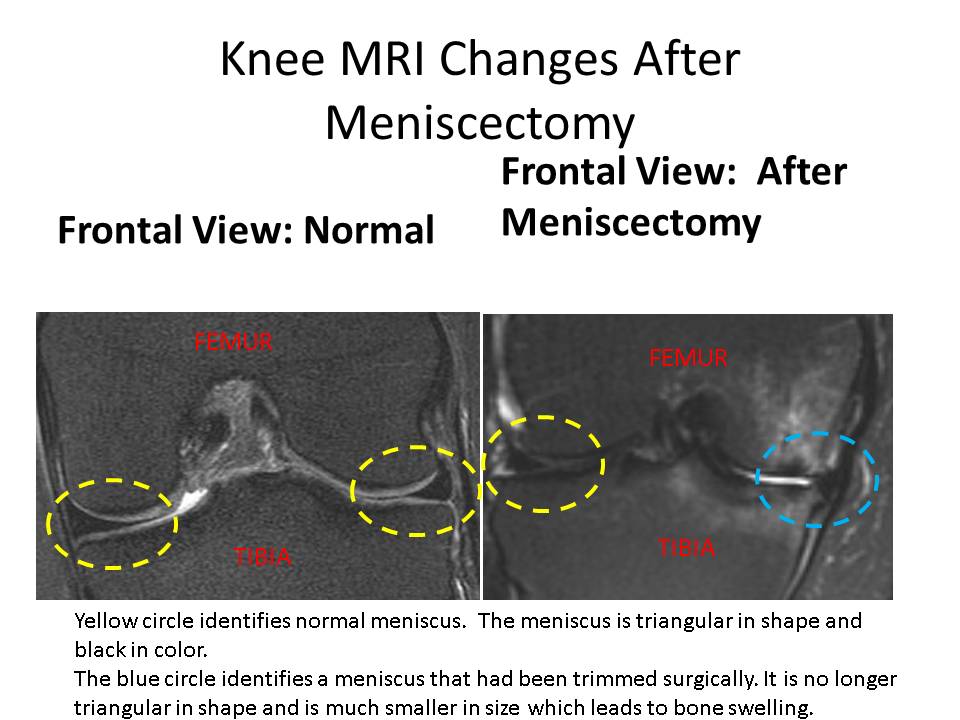
The meniscus is changed in size after a meniscectomy. That means that there is more force on a smaller surface area. The force arises from daily activities such as walking and running. This increased force in combination with changes in architecture following the surgery led to degeneration. Often times the mensicus is also displaced from the joint space. In the end you have a shock abosber that is smaller in size, prone to weakness and degeneration and partially pushed out of the joint space. The result is a nonfunctional shock absorber which exposes the cartilage surfaces to increased force. Over time the increased force leads to bone swelling (edema) which has as patchy white appearance on T2 MRI images.
MRI changes following knee meniscectomy are illustrated below. The image on the left is a normal knee MRI. Normal meniscus are triangular in shape and black in color. They are outlined in yellow circles. The MRI on the right is after meniscectomy and significant for a change in the size and shape of the mensicus which is identified by blue circle. A smaller mensicus in this case failed to cushion the thin bone from the shin bone resulting in bone swelling.

