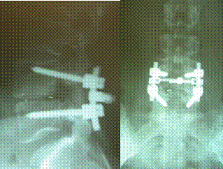
In a previous blog, I have discussed the limitations of MRI’s in identifying the source of a given patient’s pain. A clinical evaluation today illustrates this point.
35y/o athletic patient presented with a 4 year history of lower back pain, constant in duration, 6/10 in severity, progressive in nature, localized in left lower back and buttock area without any radiations into his leg. Pain which was aching in character was aggravated by prolonged sitting, standing and twisting.
Treatment to date had included massage, chiropractic care, physical therapy, trial of anti-inflammatory agents, narcotics and a surgical evaluation.
Physical examination was significant for tenderness in the left lower spine and buttocks with no neurologic abnormalities. Direct pressure applied to the mid-buttock was painful.
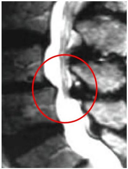
MRI of the lumbar spine was significant for advanced degeneration of the L5/S1 disc and bone swelling.
The patient was convinced that his pain was arising from the degenerative lumbar disc. Family members, his primary care physician and surgeon endorsed this view. The surgeon had recommended lumbar fusion to relieve his pain.

Low back pain can arise from many structures including muscle, ligament, facet, disc and sacro-illac joint (SI). Evaluation to determine the source of the pain had not been performed. At the Centeno-Schultz Clinic this is achieved by injecting a small volume of local anesthetic under x-ray into a specific targeted tissue. If the pain is significantly relieved from the injection, the pain generator has been identified and an appropriate treatment plan can be created. Often without such diagnostic evaluations, the source of a given patient’s pain cannot be established and therefore the patient is at risk for incorrect diagnosis and therapy.

