The headache persists despite all the different treatments and doctors. Your head feels heavy like you have a bowling ball on top of your shoulders. What is the Transverse Ligament? What is the function of the Transverse ligament? Where is the transverse ligament? What does craniocervical instability feel like? What are the treatment options for craniocervical instability? Let’s dig in.
What Is the Transverse Ligament?
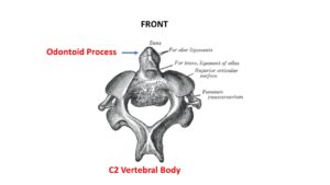
A brief review of anatomy and terminology is helpful. A ligament is a thick band of connective tissue that connects one bone to another. The cervical spine is compromised of 7 vertebral bodies which are the building blocks of the spine. They are numbered from 1-7. A “C” precedes each number to indicate that it referencing the cervical spine as opposed to the low back or thoracic spine. The C1 vertebral body connects with the head above and the C2 vertebral body below. The C2 vertebral body is unusually shaped as it has a vertical boney mass that resembles the Washington monument called the odontoid process. The odontoid process extends upward from the C2 vertebral body to reach almost the top of the C1 vertebral body (1). The transverse ligament is a ligament that resembles a seat belt that fits snugly across the base of the odontoid process.
What Is the Function of the Transverse Ligament?
Acting as a seat belt that snugly fits across the odontoid process, the transverse ligament keeps the odontoid process in place and stabilizes the upper cervical spine (2). Why is this important? Immediately behind the transverse ligament and odontoid process is a canal that extends the entire length of the spine. It is called the spinal canal and it contains the spinal cord, spinal nerves, and membranes. If the transverse ligament is damaged or is loose due to degeneration or genetic disorders, upper cervical instability can ensue. High energy trauma is the most common cause (3). Instability of the upper cervical spine can lead to irritation or compression of the spinal canal, membranes, exiting nerves, and or spinal cord. This condition is called craniocervical instability (CCI) (4). To learn more about craniocervical instability, it’s symptoms and treatment, please visit the CCJ Instability Institute
Where Is the Transverse Ligament Located?
The transverse ligament is located at the first cervical vertebral body. It acts like a seat belt and snugly holds a boney process of the second cervical vertebral body (C2) that resembles the Washingon Monument. The boney process is called the odontoid process.
What Does Cervical Instability Feel Like?
Symptoms of upper cervical instability vary depending upon the severity of the instability. The most common symptoms associated with Craniocervical Instability (CCI) include:
Headache: Debilitating in character which can be constant in duration making simple tasks and activities almost impossible
Dizziness: Simple activities such as walking down a hall can be a challenge. Many patients are consequently confined to their bed or sofa (5)
Brain Fog: Mental acuity and execution of tasks are severely impaired. Full-time employment is almost impossible for most patients
Could Craniocervical Instability Be the Cause of Your Symptoms?
Step 1
Imaging-Get movement based imaging
(DMX or Upright MRI with flexion and extension).
Step 2
Get Typed via a Telemed Appointment-There are 8 different types of CCI based on which
ligaments are Injured.
Step 3
Exam-If you’re a candidate for precise orthobiologic injections, you fly in for an exam and
treatment. A hands-on exam refines what we need to treat.
Step 4
Treatment-Get treated with a CCI focused, orthobiologic treatment plan tailored to you. That
includes PICL or whichever image guided, precise injections are the most likely to help you.
Treatment is based upon the severity of the instability and symptoms. When possible first-line treatment should be conservative in nature and include stabilization. Specialized chiropractic care is oftentimes helpful. In cases unresponsive to conservative care or severe in nature, surgery with a fusion between the base of the skull and the cervical spine has been the standard of care. The goal is to provide stabilization.
But…
In 2015 the Centeno-Schultz Clinic pioneered a nonsurgical treatment for patients with CCI. This involves the injection of a patient’s own stem cells into the damaged or loose transverse ligaments. The procedure is only performed at the Centeno-Schultz clinic and to date, we have completed over 230 cases. The results have been transformative for many which I have discussed in an earlier blog. To learn more please watch the video below.
In the neck 7 building blocks are stacked upon one another. They are called vertebral bodies. They are numbered C1-7. C2 has a unique vertical boney mass that extends up to the top of the C1 vertebrae body. It resembles the Washington Monument and is called the odontoid process. The transverse ligament wraps around the odontoid acting like a seat belt. It provides important stability for the C1/2 joint and upper cervical spine. Injury to the transverse ligament can occur due to trauma or genetic disorders. This can lead to irritation or compression of the spinal canal, membranes, exiting nerves, and or spinal cord. The condition is called craniocervical instability. Symptoms include headache, dizziness and brain fog. A breakthrough occurred in 2015 when the Centeno Schultz Clinic pioneered a nonsurgical treatment for transverse and alar ligament injuries. The procedure called a PICL uses a patient’s own bone marrow-derived stem cells to accelerate the healing of the damaged cervical ligaments.
Has trauma resulted in a dramatic change in your level of function? Have your new headaches and dizziness baffled your traditional doctors? Are all your radiographic studies been “normal”. Schedule a Telemedicine evaluation with a board-certified, fellowship-trained physician who can review your history, imaging, and determine your best treatment options. Stop the suffering and schedule an evaluation today.