Hey, it’s Dr. Centeno. And what is an Upright MRI? And can it diagnose CCJ instability? So, CCJ Instability means that the upper neck bones move around too much due to lax ligaments. Lots of different ligaments up there that can be torn, partially torn, stretched, or just loose due to other problems.
And there are two types of imaging that can suggest CCJ Instability. One is static or not moving imaging and the other is dynamic. And in the case of upright MRI, that’s generally a combination of static and dynamic.
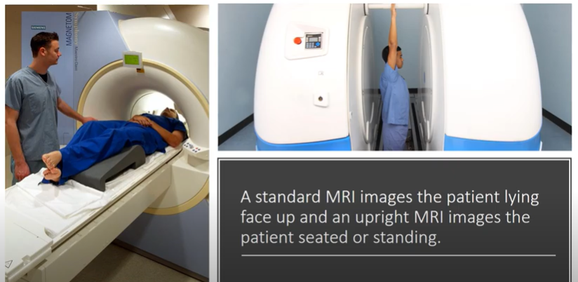
So, standard MRI images like on the left here are done lying face up or what we call supine; an upright MRI images the patient seated or standing (see Figure 1, below). Now, there are obviously advantages to this because one is weight-bearing, how you would normally exist on a day-to-day basis and the other is not.
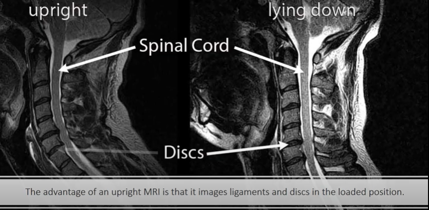
An advantage of upright MRI is that it images the ligaments and discs in the loaded position. So you can see here (see Figure 2, below), the MRI on the right lying down or what we call supine doesn’t show as much spinal cord compression as the one upright.
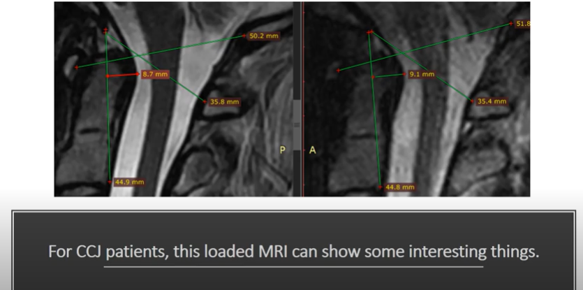
For CCJ patients, this loaded MRI can have some interesting things. If we just look at a static measurement, the one on the left was taken lying face up. The one on the right was taken upright (see Figure 3, below). And the one on the right shows a little bit more abnormal values than the one on the left. Some of the values on the left are normal and some of them by the time the patient is seated or standing and imaged become abnormal.
Our research group published a paper on Chiari malformation, which is low-hanging cerebellum which is part of the brain, and upright imaging and found that this particular problem could be found better on upright imaging. So interesting stuff.
The downside to upright imaging is that the imaging resolution is often lower due to the lower MRI field strength that accommodates the ability to have a more open bore that allows motion.
An advantage on the other hand of upright MRI is the ability to see what the structures are doing as they move. So we have a patient here on the left that is flexing her neck and on the right that’s extending the neck. Again, we can see some things buckling between these two that we wouldn’t normally see on a supine MRI.
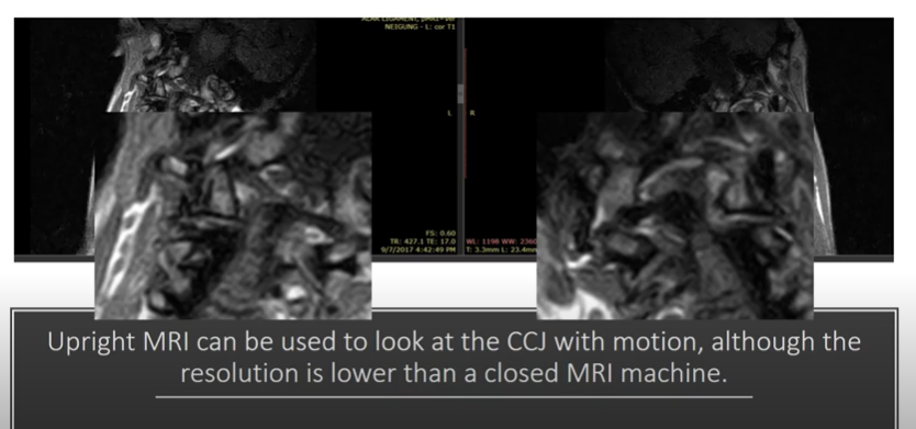
An Upright MRI can be used to look at the CCJ with motion, although again, the resolution is lower. So here (see Figure 4, below) we’ve got lateral bending looking at the alar ligaments. And you can see the alar ligaments, the resolution isn’t great, but you can get some idea of what happens to the relationship between C1 and C2.
So is upright and dynamic MRI or supine which is lying face up in a tube and static MRI imaging right for you in trying to diagnose what’s going on with your upper neck? Well, that’s a decision we can help you with at the CCJ Instability Institute. So go to ccjinstability.com to learn more. Thanks so much for watching and have a great day.
Could Craniocervical Junction (CCJ) Be The Cause Of Your Symptoms?

Imaging-Get movement based imaging (DMX or Upright MRI with flexion and extension).

Get Typed via a Telemed Appointment-There are 8 different types of CCJ based on which ligaments are Injured.

Exam-If you’re a candidate for precise orthobiologic injections, you fly in for an exam and treatment. A hands-on exam refines what we need to treat.

Treatment-Get treated with a CCJ focused, orthobiologic treatment plan tailored to you. That includes PICL or whichever image guided, precise injections are the most likely to help you.