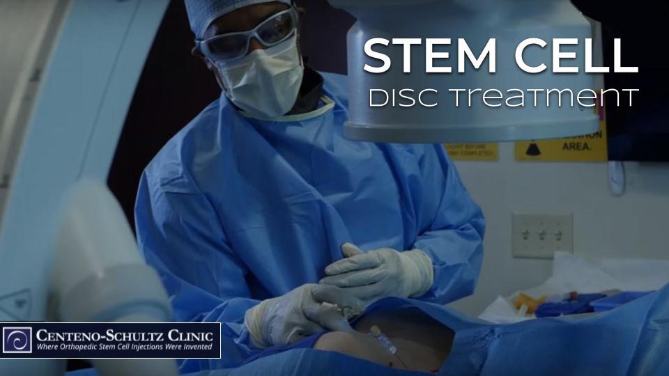
Today, we invite you into one of our interventional orthopedics procedure suites. Dr. Pitts will be performing an advanced image-guided stem cell injection into an L4–5 disc to treat a patient with an annular tear and low back pain. Watch the video below, and you can also review terminology and equipment in this post below the video.
Terminology for the Stem Cell Disc Treatment
Dr. Pitts does a fantastic job of narrating his procedure as he goes, but there is some medical terminology we will define below to help you follow along.
Annular tear: Each disc has an outer covering called the annulus. A tear can occur in this outer covering, and this is called an annular tear. In the video, the patient has an annular tear in the L4–5 disc, located between the L4 and L5 vertebrae in the lumbar section of the spine. If this tear were not treated, a bulging disc can occur, which is when the gel inside the disc causes a bulge in the torn or damaged area. If it progresses to a complete tear, a herniated disc can result, which is when the gel inside squirts out. Back fusions are common for disc issues, but we do not recommend this surgery as it can cause adjacent segment disease and other problems.
Contrast Dye: The substance being injected (termed the injectate) is invisible on X-ray. Contrast dye is injected as well as it can be seen on X-ray, and it confirms that the injectate is making it to its intended location. In this particular case, the contrast also allows visualization of the annular tear as you will see on the video. Dr. Pitts is then able to inject the orthobiologic into the tear.
Extravagate: This terminology describes the contrast leaking outside the limits of the normal interior disc space. If the disc is healthy, the contrast will stay in the nucleus (middle) of the disc; if the disc is torn, the contrast will leak out (extravagate) of the nucleus and into the damaged area. In this patient, you will see the contrast extravagate, identifying the annular tear.
Intradiscal: This terminology simply means inside the disc. An intradiscal procedure is one that takes place within the disc.
AP view: This is an X-ray view of a structure taken from an anteroposterior (front-to-back) position. In this case, the AP view is of the L4–5 disc. After the contrast is injected, the AP view is observed, which confirms proper needle placement and good contrast flow within the disc.
Lateral view: This is X-ray view of a structure taken from a side position. Again, in this case, the lateral view is of the L4–5 disc. This is a second step that confirms proper needle placement. In this case, this is the view that allows Dr. Pitts to observe the contrast leaking out of the middle of the disc. This confirms there is an annular tear, and he is then able to perform the precise injection of the orthobiologic.
Equipment You Will Observe in the Video
Fluoroscopy imaging is a live X-ray where Dr. Pitts can visualize in real-time everything he is doing, such as determining precise placement of the needle. You will also see some of our onsite lab equipment in our state-of-the-art lab and a lab technician processing cells. For comparison, you’ll also get a peek at a one-size-fits-all automated bedside machine; if your provider is using one of these to concentrate your cells, he or she is not customizing the stem cell treatment precisely for you.
You’ll see a full surgical monitor that measures vital signs throughout the procedure, and we always have emergency equipment (e.g., oxygen, crash cart, automated defibrillator, etc.) on hand. If you want to learn more about our procedure-suite setup, click here and scroll to the second half of the post.
Why Regenexx Stem Cell Disc Treatments Are Very Different
Most providers who perform stem cell disc treatments do it blindly, meaning without imaging guidance to determine precise needle placement. Blind injections mean most of the cells end up in the muscles rather than the disc, as there’s no way to confirm proper placement in any specific structure.
For those who do use guidance, what separates our Regenexx intradiscal treatments from other stem cell providers performing the procedure is our ability to customize the treatment to each patient. This is because we process everything by hand in our onsite lab. Stem cell disc treatments require a high concentration of stem cells in a minuscule volume. Why? Discs cannot hold a lot. And our treatment allow for this smaller customization. With that one-size-fits-all automated bedside machine, there tends to be a smaller number of stem cells in a higher volume of injectate. The result? A significantly lower concentration of cells injected into the disc.
With a disc treatment it’s imperative to perform the right type of procedure for the right type of disc problem. This could be a platelet procedure to tighten up loose ligaments around a disc. It could be injecting platelets into damaged facet joints. It rarer cases, it could be a stem cell disc treatment for an annular tear that requires a stem cell injection into the disc itself. Whatever your disc issue, the treatment is customized to the individual patient and issue. We hope you’ve enjoyed this visit to our procedure suite with Dr. Pitts.

