Thoracic Spine Osteophytes
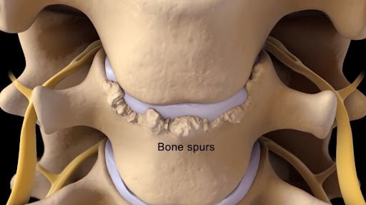
An osteophyte, also known as a bone spur, is an abnormal bone growth that occurs at the edge of a bone. Thoracic spine osteophytes are bone spurs that occur in the thoracic spine. The thoracic spine is that part of the spine located below the neck and above the lower back. It is often referred to as the mid back.
There are many causes of thoracic spine osteophytes. The major cause is instability. A bone spur in most cases is simply the body’s response to an unstable environment.
What Are Osteophytes in the Thoracic Spine?

Osteophytes are abnormal bone growths that typically occur at the edge or end of a bone, near a joint. They can also occur in the cervical, thoracic, or lumbar spines. In the thoracic spine, they can occur in many different places. The most common locations for bone spurs include:
- Central spinal canal
- Neuroforamen
- Facet joint
- Costotransverse joint
- Within a ligament
Symptoms Associated with Osteophytes in the Thoracic Spine
The symptoms associated with osteophytes in the thoracic spine will vary depending upon multiple factors that include:
- Location of the thoracic bone spur
- Size
- Whether it is compressing or irritating adjacent tissue
- Presence or absence of spinal cord or exiting nerve irritation/injury
Stiffness
Bone spurs can impinge on the normal movement of the spine. As the spine is a highly flexible structure, any obstruction to its movement can lead to stiffness.
Inflammation may also play a role. Bone spurs can cause inflammation in the local tissues. Inflammation, in turn, can cause swelling and irritation, contributing to stiffness.
Reduced Range of Motion
Bone spurs may limit forward, backward, or side-to-side movements, making them painful and thereby reducing the range of motion.
Muscle Weakness
Muscle weakness may occur if a bone spur in the thoracic spine is located within the central spinal canal or neuroforamen. When in the central spinal canal, an osteophyte can compress or damage the spinal cord, creating several potential neurologic symptoms, which include muscle weakness.
The neuroforamen is the bony doorway through which nerves exit the spine. There is a right and left neuroforamen at each level of the spine. An osteophyte within or near the neuroforamen can irritate or compress the exiting nerve, leading to muscle weakness. In severe cases, it can lead to muscle shrinkage (atrophy).
Pain When Breathing
Breathing involves two principal actions:
- Inspiration: drawing air into the lungs
- Expiration: expelling oxygen from the lungs
Inspiration and expiration both require movement of the chest wall. With inspiration, the chest wall expands, lifting the ribs and drawing the oxygen in. Conversely, expiration involves contraction of the chest wall, thereby forcing the oxygen out.
Chest wall expansion and contraction can cause thoracic spine bone spurs to irritate nerves or adjacent tissue, making breathing painful.
Nerve Pain in the Thoracic Spine
The thoracic spine is the part of the spine below the neck (cervical spine) and above the low back (lumbar spine). It is often referred to as the mid back. Nerves exit the thoracic spine at each level and can become irritated, compressed or injured, resulting in pain and dysfunction. This is commonly referred to as thoracic radiculopathy or pinched nerve.
Read More About Nerve Pain in the Thoracic SpineThoracic Spine Pain
Simply put thoracic spine pain is pain that arises from the thoracic spine. It may be acute or chronic. It may be constant or intermittent. It may be mild or can be so severe as to take your breath away. To better understand thoracic spine pain please review the sections below. The thoracic spine is that part of the spine that is sandwiched between the neck and low back. Many refer to it as the middle section of your spine. It starts at the base of your neck and ends at the bottom of your ribs. The thoracic spine is the longest region in the spine.
Read More About Thoracic Spine PainCauses of Osteophytes in the Thoracic Spine
Osteophytes in the thoracic spine can occur due to various factors. Understanding the underlying causes of thoracic osteophytes is crucial for effective diagnosis and treatment strategies.
Aging
The aging process is a significant factor in the development of osteophytes in the thoracic spine.
As we age, our discs typically lose water content and degenerate, leading to changes in the structure and stability of the spine. This degeneration and instability can contribute to the formation of bone spurs.
Degenerative Disc Disease
Degeneration of the thoracic spine discs can lead to the formation of osteophytes. Degenerative changes include loss of disc height and water content. This may compromise the disc’s ability to absorb the forces of daily living, leading to facet joint overload and spine instability, both of which can stimulate the formation of bone spurs.
Osteoarthritis
Osteoarthritis is common in the thoracic spine and involves the breakdown of cartilage in the facet, costovertebral, and costotransverse joints. Cartilage breakdown can compromise the joint, its capsules, and its stability, leading to the formation of bone spurs.
Spinal Injuries
Prior injuries to the thoracic spine, such as fractures, dislocations, or subluxations, can cause instability and abnormal stress on the spinal joints and discs. This can lead to the development of osteophytes as the body tries to repair and stabilize the injured area.
Ligament Instability
Ligaments are thick pieces of connective tissue that connect bone to bone. They provide significant stability for the spine. Ligaments are susceptible to injury and degeneration, which in turn can compromise spinal stability. One of the ways the body attempts to correct spinal instability is through the formation of osteophytes.
Listhesis
Listhesis is a medical condition in which a vertebral body moves in relation to the vertebrae above and below it. If it slips forward, this condition is referred to as an anterolisthesis, whereas if it slips backward it is referred to as retrolisthesis. Listhesis is an unstable condition and in many cases can lead to the formation of osteophytes in the thoracic spine.
Spinal Stenosis
Thoracic spinal stenosis is a medical condition characterized by a narrowing of the spinal canal, causing potential irritation or injury to the spinal cord and nerves. The increased forces caused by spinal stenosis can trigger the formation of bone spurs.
How Are Thoracic Osteophytes Diagnosed?
Thoracic osteophytes, or bone spurs in the thoracic spine, are typically diagnosed through imaging studies. Imaging studies are commonly ordered after a patient has undergone a thorough history and physical examination. The most common radiographic studies include:
- X-ray: X-rays are often the initial imaging test used to visualize the bones and assess for the presence of an osteophyte. X-rays are affordable and readily available in most communities. They can show bony changes such as bone spurs as well as fractures. X-rays do have limitations as they cannot show soft tissue.
- MRI (magnetic resonance imaging): MRI is an imaging modality that uses strong magnetic fields and radio waves to generate detailed images of the body. It can provide detailed images of soft tissue structures such as nerves, spinal cord, blood vessels, and the brain. MRI can identify thoracic spine osteophytes and whether or not the spinal cord and/or nerves are being compressed.
- CT scans (computerized tomography): CT scans provide more detailed images of the spine and can help visualize the size and location of osteophytes. CT scans are relatively quick but do involve exposure to radiation.
Common Treatment Options for Thoracic Osteophytes
Thoracic osteophytes can be painful, potentially limiting range of motion and function. The severity of the pain and the degree of compromise of movement and function in most cases will depend upon the size of the bone spur, its location, and whether there is any nerve or spinal cord irritation.
When appropriate, conservative treatment should always be first-line treatment. Common treatments include:
Prescription Pain Medications
Medications are often utilized for the pain and stiffness associated with osteophytes in the thoracic spine. Common examples include:
- Natural non-steroidal anti-inflammatory agents like fish oil and curcumin
- Pharmaceutical anti-inflammatory agents. Common examples include Celebrex, ibuprofen, and diclofenac. These are powerful anti-inflammatory agents that should be avoided, due to their significant side effects. They are available in oral tablets and topical creams.
- Muscle relaxants. Patients are often prescribed muscle relaxants when thoracic spine tightness persists despite rest and anti-inflammatory agents. Common examples include Flexeril, Skelaxin, Robaxin, and Soma.
Physical Therapy (PT)
Physical therapy can be helpful for patients with osteophytes in the thoracic spine by addressing pain and improving mobility and function. Therapeutic exercises can improve flexibility and strength. Stretching can improve the flexibility of muscles and soft tissue. Core stabilization can provide better support to the spine and improve compromised postures.
Surgery
When the pain and compromised function that is secondary to bone spurs do not respond to rest, physical therapy, or pain medication, surgery is often recommended. The most common surgeries for bone spurs are:

- Thoracic laminectomy
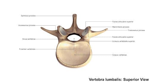
The vertebrae are the bony building blocks that stack one upon another to make up the cervical, thoracic, and lumbar spines. The vertebrae are composed of the body and the posterior arch. The posterior arch is composed of the pedicles and the laminae.
The laminae are thin flat bones that arise from pedicles and create a bony central canal. This central canal contains and protects the spinal cord and spinal nerves.
Conceptually, the laminae can be thought of as the bony roof that protects the spinal cord and nerves. A laminectomy is a surgical procedure where the lamina is removed. A hemilaminectomy is a surgical procedure where only one of the lamina is removed. Recall that there is a right and left lamina at each level of the spine. A laminectomy involves the removal of both the right and left laminae.
- Foraminectomy
The neuroforamen is a bony foramen through which nerves exit the spine. There is a right and left neuroforamen at each level of the spine. A bone spur can compromise the patency of the foramen as well as irritate or compress exiting nerves. A foraminectomy is a surgery that can be conceptualized as a rotator rooter. It removes any bone spurs, scar tissue, or other tissues that narrow the foramen or irritate or compress the exiting nerve.

- Spinal fusion
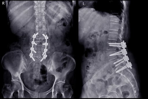
A spinal fusion is a major surgery that involves the removal of one or more discs, which are then replaced with either bone grafts or spacers. The spine is then stabilized with screws, plates, and rods. The hardware can be inserted posteriorly or anteriorly through the abdomen. There are many indications for spinal fusion, which include disc herniation.
Regenerative Options for Thoracic Osteophytes
Osteophytes in the thoracic spine typically occur as a result of some type of ligament, joint, or spinal instability. It is the body’s response to an unstable spine. Over time, additional abnormal bone is laid down in an attempt to stabilize the spine. Pain medications, including narcotics and steroids, do not address the underlying problem but simply help ease the symptoms.
At the Centeno-Schultz Clinic, we undertake a different approach that is referred to as the functional spinal unit (FSU). We acknowledge the complexity of the human body and how all the different parts work together in a highly synchronized manner. For the very best results, all the different parts of the spine must be evaluated and treated together. Treatment aims to heal the damaged or degenerative tissue responsible for a given patient’s symptoms. Treat the underlying problem and, in many cases, the symptoms improve or resolve. If you simply treat the symptoms, in many cases the underlying injury progresses along with the symptoms.
Treatment options include:
Prolotherapy Injections
It has been successful in the treatment of many disorders including neck, shoulder, knee, and ankle pain. Dr. Centeno recently published an article in The Journal of Prolotherapy in which he discusses the use of x-ray guidance with prolotherapy. This ensures that the injection is in the correct place to maximize clinical results. Dr. Centeno discusses the use of prolotherapy for the treatment of neck, knee, sacroiliac joint, ankle, ischial tuberosity, and shoulder pain. At the Centeno-Schultz Clinic x-ray guided prolotherapy is just one of the therapies utilized in the successful treatment of pain. Regenerative injection therapy (RIT) or prolotherapy…
Read More About Prolotherapy InjectionsPRP Injections
PRP is short for platelet-rich plasma, and it is autologous blood with concentrations of platelets above baseline values. The potential benefit of platelet-rich plasma has received considerable interest due to the appeal of a simple, safe, and minimally invasive method of applying growth factors. PRP treatments are a form of regenerative medicine that utilizes the blood healing factors to help the body repair itself by means of injecting PRP into the damaged tissue. In regenerative orthopedics, it is typically used for the treatment of muscle strains, tears, ligament and tendon tears, minor arthritis, and joint instability. There have been more than 30 randomized controlled trials of PRP…
Read More About PRP InjectionsGet Innovative Approaches to Managing Thoracic Osteophytes
Thoracic osteophytes, also known as bone spurs, are abnormal bone growths that are typically the result of instability. They can occur in many different parts of the thoracic spine, which include the central spinal canal, neuroforamen, facet joint, costotransverse joint, and ligaments.
Symptoms vary depending upon many different factors including the size of the spur, its location, and whether there is any irritation or compression of the adjacent tissues and nerves. Common symptoms include stiffness, reduced range of motion, muscle weakness and pain with breathing, and pain.
The most common causes of osteophytes in the thoracic spine include aging, degenerative disc disease, osteoarthritis, spinal injuries, ligament instability, listhesis, and spinal stenosis.
Osteophytes are typically diagnosed by radiographic imaging, which can include X-rays, CT scans, and MRI. MRI is a powerful imaging modality as it can visualize soft tissue such as ligaments, tendons, nerves, spinal cord, spinal fluid, and blood vessels.
Common treatment options include PT and pain medications. When these fail to provide significant benefits, patients may be referred for surgical consultation.
Bone spurs are commonly the result of some instability. Utilizing the functional spine unit approach, the physicians at the Centeno Schultz Clinic can identify the actual source of the instability and direct treatment toward it. Regenerative treatment options include PRP and prolotherapy, which can help heal the underlying problem and instability.
Thoracic spine osteophytes, aka bone spurs, are a good indication that some form of instability exists in the thoracic spine. If left untreated, the instability along with the bone spur may increase in size. Ignoring the problem can lead to an increase in the instability, symptoms, and size of the bone spur, thereby limiting less invasive, nonsurgical options.
If you or a loved one suffers from thoracic spine osteophytes, please consider an in-office or virtual evaluation with one of the physicians at the Centeno-Schultz Clinic. Learn about regenerative treatment that may be right for you and stop the suffering.
Are you a Candidate?

John Schultz, MD
John R. Schultz M.D. is a national expert and specialist in Interventional Orthopedics and the clinical use of bone marrow concentrate and PRP for orthopedic injuries. He is board certified in Anesthesiology and Pain Medicine and underwent fellowship training. Dr. Schultz has extensive experience with same day as well as culture expanded bone marrow concentrate and sees patients at the CSC Broomfield, Colorado Clinic, as well the Regenexx Clinic in Grand Cayman. Dr. Schultz emphasis is on the evaluation and treatment of thoracic and cervical disc, facet, nerve, and ligament injuries including the non-surgical treatment of Craniocervical instability (CCI).
Other Resources for Thoracic Spine Osteophytes
-
What Happens If You Have Back Pain From Golf?
Back pain is a common complaint among golfers, impacting both amateur enthusiasts and professional athletes. Golf, while seemingly low-impact, involves repetitive, high-intensity movements that can stress the spine and surrounding structures. Understanding the causes, symptoms, and preventative measures for golf-related back pain can help maintain performance and long-term health. Golf And Back Pain The golf…
-
Thoracic Spinal Fusion Recovery Explored
Thoracic spinal fusion is an intricate surgery that involves fusing vertebrae to reduce pain and improve functionality. It is often necessary due to conditions such as scoliosis, spinal fractures, or degenerative disc disease. Recovery from thoracic spinal fusion is a critical phase, requiring careful management to ensure proper healing and restoration of movement. Patients must…
-
The C1 And C2 Vertebrae – What To Know
The C1 and C2 vertebrae, also known as the atlas and axis, are the uppermost bones in the spinal column. They play a crucial role in supporting the skull and enabling head movements. Damage or injury to these vertebrae often leads to pain, limitations to daily activities, and reduced quality of life. Many patients, without…
-
Spinal Fusion Complications Years Later – What You Should Know
Spinal fusion surgery, often performed to alleviate chronic back pain or spinal instability, can lead to complications years later. These may include adjacent segment disease, where nearby vertebrae deteriorate, chronic pain, or hardware failure. Patients might also experience limited mobility and nerve damage over time. Understanding these potential risks is crucial for long-term care and…
-
Spinal Fusion Recovery: What to Expect
Navigating spinal fusion recovery can be a daunting prospect, given its impact on daily life and mobility. Understanding what to expect during this process is crucial for individuals undergoing this procedure. In this article, we’ll explore the typical timeline, challenges, and strategies for managing recovery after spinal fusion surgery, providing insights to help individuals prepare…
-
Back Cracking: The Truth of What’s Actually Happening in Your Body
Back cracking is a phenomenon that many people experience, often eliciting both curiosity and concern. Whether it’s the satisfying pop from a morning stretch or the deliberate twist during a yoga session, the sound and sensation of cracking your back can be oddly gratifying. But what exactly is happening inside your body when you hear…
-
Back Fusion
Spinal fusion, also known as back fusion, is a surgical procedure designed to help severe spinal instability that causes severe pain or nerve injuries. It involves permanently connecting two or more vertebrae in your spine to eliminate motion between them. This article will delve into the intricacies of spinal fusion, exploring the reasons behind the…
-
Understanding the Normal Curvature of the Spine
The human spine, a marvel of engineering, is not a straight column but rather a structure with gentle curves. These natural curves are essential for maintaining balance, allowing flexibility, and absorbing the shock of movement. The spine’s curvature plays a critical role in overall health, influencing posture, mobility, and the function of the nervous system. …
-
Understanding the Thoracic and Lumbar Spines
The thoracic spine and lumbar spine make up a vital nexus of stability and mobility in the human body. In this exploration, we delve into the biomechanics and complexities that define these regions, unraveling their significance in posture, movement, and overall well-being. Understanding the thoracic and lumbar spine not only illustrates the mechanics of our…