The headaches, brain fog and dizziness were getting worse and unresponsive to conservative care. You were referred for surgical evaluation. During the consultation time was spent reviewing your neck MRI. Different measuresurements were performed. Should you be concerned? What is the Cranium? What is the Craniocervical junction (CCJ)? What is the Grabb Oakes Measurement? How is it determined? What is a normal Grabb Oakes Measurement? Why is it important? Let’s dig in.
Introduction
The Grabb Oakes Measurement is a radiographic tool used in the evalution of upper neck and brain disorders.
Upper Cervical Anatomy Tidbits
Understanding the anatomy is central to understanding how to determine the Grabb Oakes Measurement. Here are some key points.
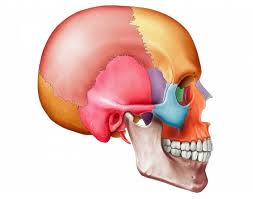
What Is the Cranium?
The Cranium is your skull. Otherwise, known as your noggin. The brain is contained and protected by the Cranium. The brain has many different parts. The lowest part of the brain is called the Brainstem.
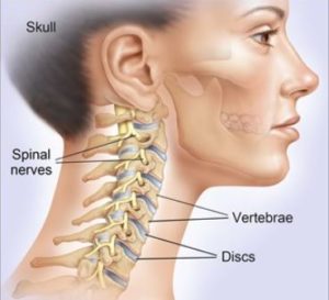
What Is the Cervical Spine
Your neck is your cervial spine. It is composed of multiple structures that include bones, discs, facet joints, ligaments, tendons, nerves and blood vessels. The bones in the neck are also known as vertebral bodies. There are 7 vertebral bodies in the neck that are stacked upon one another. They are numbered 1 through 7. The letter C precedes each number indicating that we are referencing the Cervical Spine. The top bone is the C1. Immediately underneath the C1 is the C2 bone.
What Is the Craniocervical Junction (CCJ)?
The Craniocervical Junction is the area between the Skull and the Cervical spine. It consists of the bones that forms the base of the Skull, the first two bones in the spine, and the neural structures which include the Brainstem. At the base of the skull is a large opening that is called the Foramen Magnum. Foramen meanings opening. Magnum means large. So the Foramen Magnum is a large boney opening at the base of the Skull that allows important structures to pass through. The most anterior aspect of the Foramen Magnum is called the Basion.
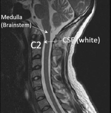
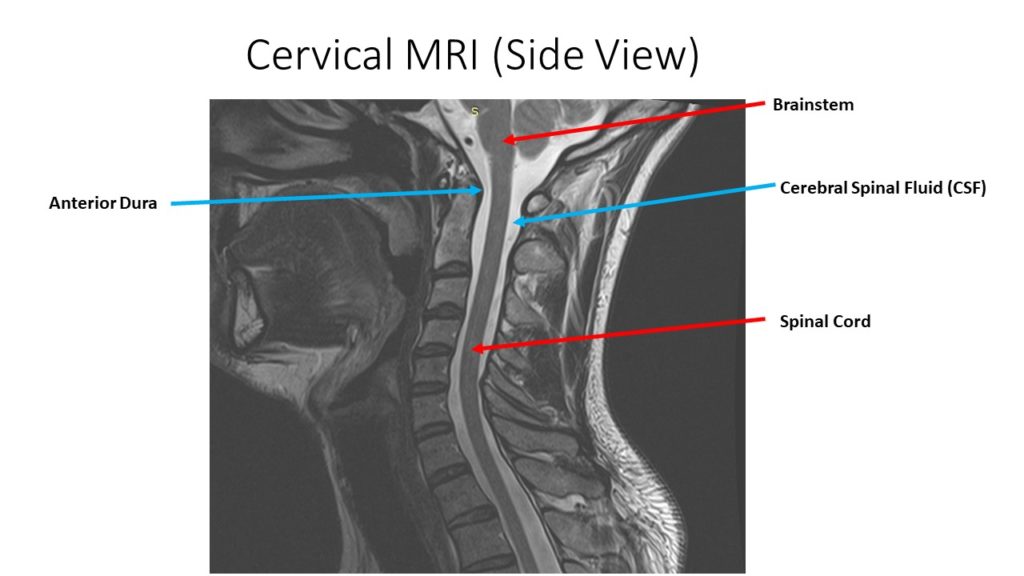
The Brainstem is the lowest part of the brain. It connects directly to the spinal cord. The Brainstem is protected and encircled by the Cerebral Spinal Fluid (CSF). This is illustrated in the side view MRI of the brain and neck shown below. The Brainstem is dark in color and located at the base of the skull. It connects directly with the spinal cord which is also dark in color. In contrast, the Cerebral Spinal Fluid (CSF) is white in color and that provides an important layer of protection. The C2 bone lies in front of the spinal cord and CSF and is identified.
Could Craniocervical Instability Be the Cause of Your Symptoms?
Step 1
Imaging-Get movement based imaging
(DMX or Upright MRI with flexion and extension).
Step 2
Get Typed via a Telemed Appointment-There are 8 different types of CCI based on which
ligaments are Injured.
Step 3
Exam-If you’re a candidate for precise orthobiologic injections, you fly in for an exam and
treatment. A hands-on exam refines what we need to treat.
Step 4
Treatment-Get treated with a CCI focused, orthobiologic treatment plan tailored to you. That
includes PICL or whichever image guided, precise injections are the most likely to help you.
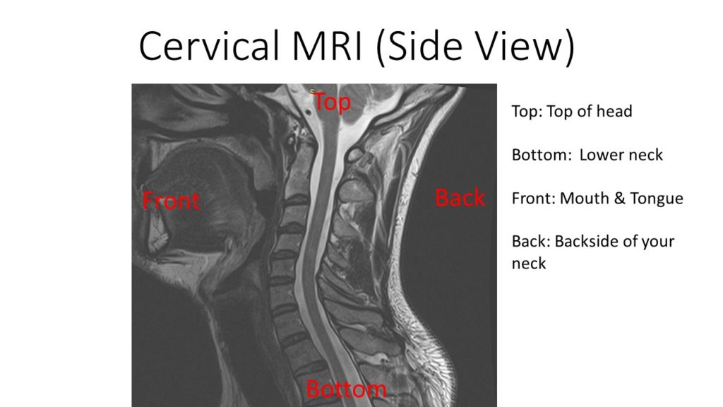
Secure Sagittal images of cervical MRI. Sagitial means side view.Note the orientation: Side View Orientation: Top. Bottom. Front (Anterior). Back (Posterior)
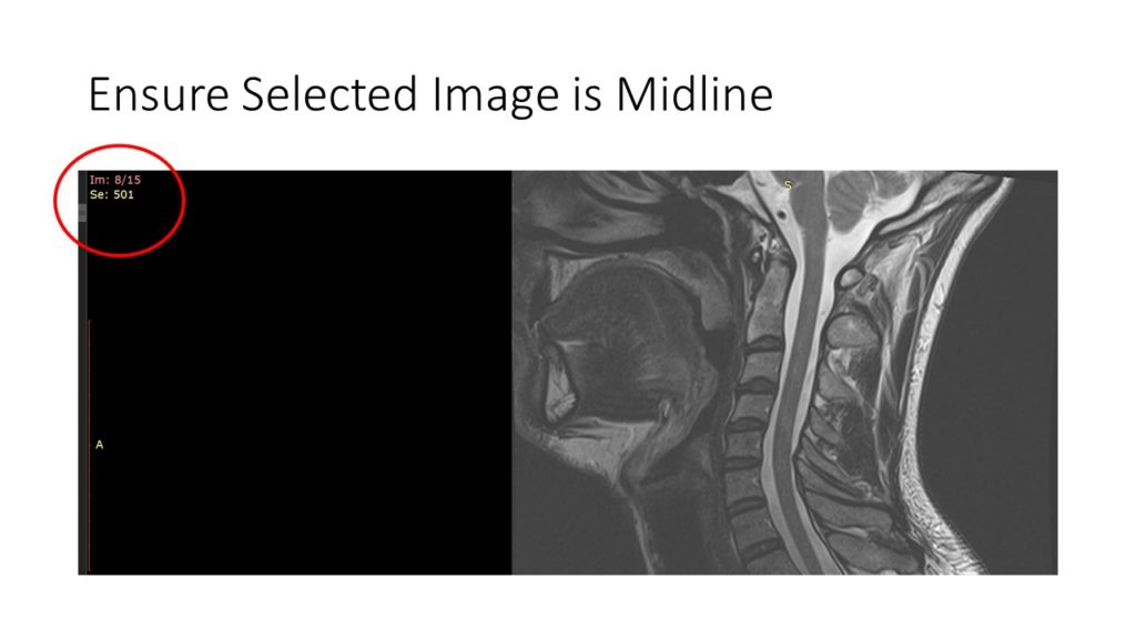
Ensure that the Sagittal image is centered.It is important that you select an image that represents the center of the spine. This can be achieved by looking at the image number that is circled in red. An MRI is simply a series of thin slices from one side of the body to other. In this particular case there are 15 images in total. Hence image 7 or 8 represents the center.
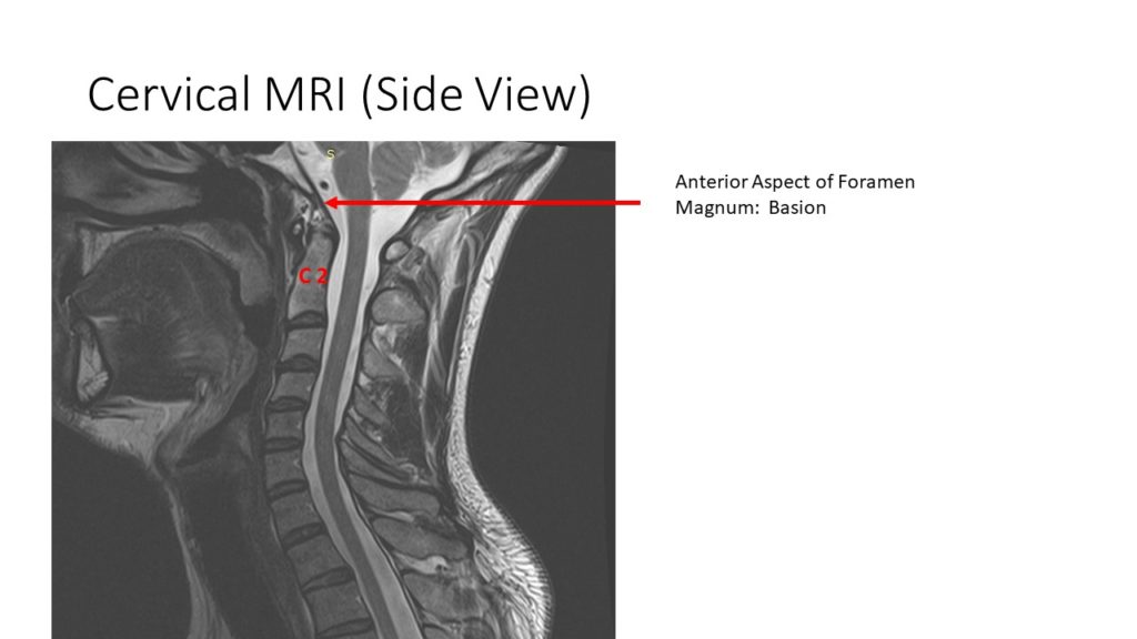
Identify the Basion.The Basion is the tip of the bone identified by the red arrow. It is a bone and represents the front aspect of the Foramen Magnum. The Foramen Magnum is the hole at the base of the skull that allows the Brainstem to pass through the Cranium and connect with spinal cord.
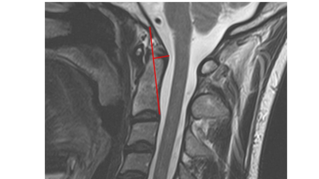
Identify the C2 boneThe C2 bone on side view looks like the Washington Monument: Tall and slender.
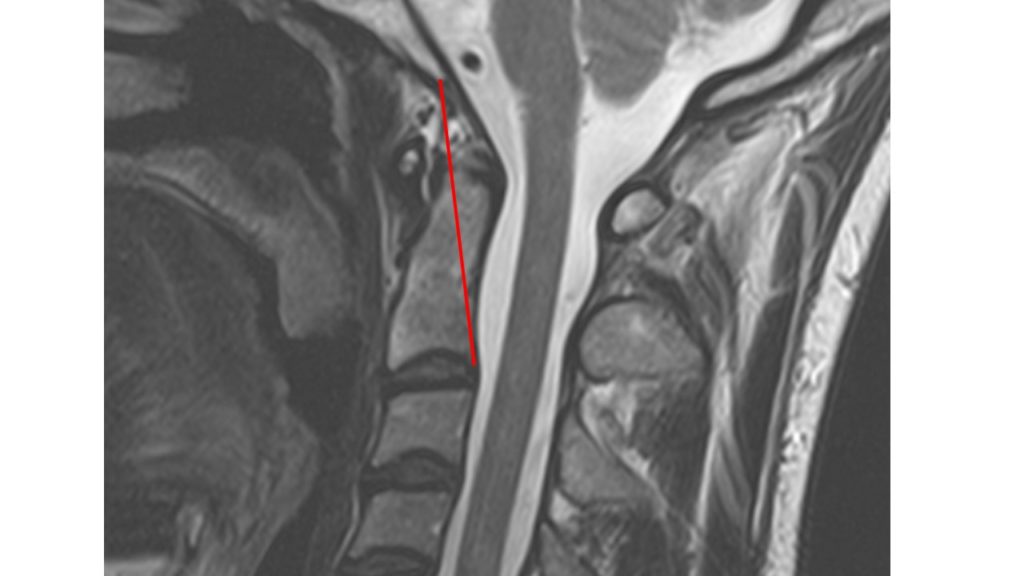
Draw a line from the Basion to the inferior(bottom), posterior aspect of the C2 bone.
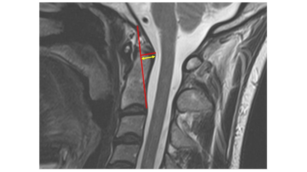
Draw a peripendicular line to the Dura.The Dura is a thick protective layer that covers the spinal cord and brain. It is the black line immediately adjacent the backside of the C2 body.
Measure the distance from the intersection of the two lines and the Dura.With a radiographic tool you measure the length of the yellow line. This is the Grabb Oakes Measurement. In this particular case it is 8.5mm.
What Is a Normal Grabb Oakes Measurement?
A normal Grabb Oakes Measurement is 9mm or less (1). It is one of many radiographic tools utilized in the evaluation of head and upper cervical spine disorders. There are a number of important structures in the upper cervical spine that are susceptible to irritation or injury that are identifed below. A future blog will discuss this topic in greater detail.
In Conclusion
The Cranium is the skull which provides a protective cover for your brain.
The Cervical Spine is composed of seven boney building blocks that stack upon one another. They are numbered from C1 to C7.
The Foramen Magnum is a large opening at the base of the skull that allows the neural elements to exit from Cranium and into the Spinal Canal. The anterior border of the Foramen Magnum is called the Basion.
The Grabb Oakes Measurement is a useful radiographic tool utilized in the evaluation of brain and upper neck disorders.
It is determined by drawing a diagonal line from the Basion to the posterior, inferior aspect of the C2 bone. A line perpendicular to this is then created extending to the Dura. The distance of this line is the Grabb Oakes Measurement.
The Centeno-Schultz Clinic are experts in the nonsurgical treatment of upper cervical injuries including Craniocervial Instability and Atlantoaxial Instability. The Grabb Oakes Measurement is one of the many tools utilized in the evaluation of patients. Treatment options include x-ray guided PRP and Bone Marrow Concentrate.
If you or a loved one have ongoing neck pain, headache, and dizziness that has not responded to conservative treatment, please schedule a telephone Candidacy discussion with a board-certified, fellowship-trained physician. At the Centeno-Schultz Clinic, we are experts in the evaluation and treatment of upper neck injuries. From the comfort of your home or office learn what treatment options are available for you.
1. Henderson FC Sr, Francomano CA, Koby M, Tuchman K, Adcock J, Patel S. Cervical medullary syndrome secondary to craniocervical instability and ventral brainstem compression in hereditary hypermobility connective tissue disorders: 5-year follow-up after craniocervical reduction, fusion, and stabilization. Neurosurg Rev. 2019;42(4):915-936. doi:10.1007/s10143-018-01070-4