At the Centeno-Schultz Clinic, we utilize the SANS approach in our evaluation and treatment of patients. This has been discussed in prior blogs.
S stands for stability. Stability is dependent upon muscle strength and ligament stability.
The Multifidus Anatomy

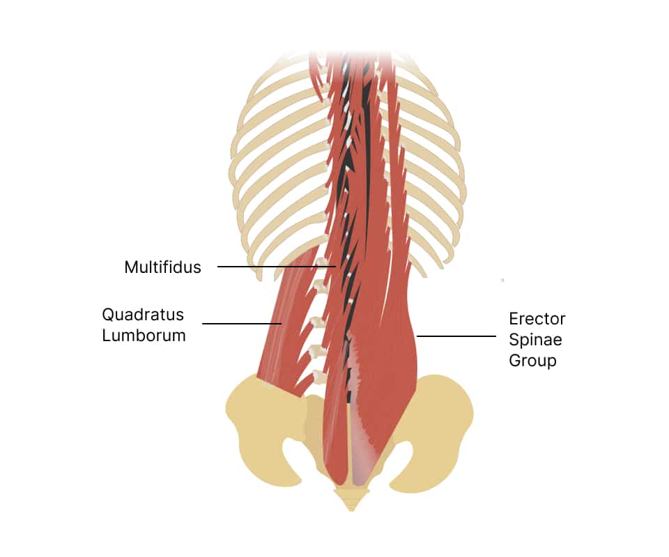
The multifidus is one of the deep muscles of the back. During development, the muscles grow towards the tailbone bone, and so the fibers run from the head to tailbone or craniocaudally. It spans about 2-4 segments. It has multiple origins which include the sacrum and ilium, the transverse processes of T1-L5, and the articular processes of C4-C7.
It gets inserted into the spinous processes of the vertebrae about 2 to 4 segments above its origin. It is a short triangle-shaped muscle that together with the deeper muscles, semispinalis, and rotatores, forms the transversospinal deep back muscle group.
A Critical Spine Stabilizer
An important stabilizer of the spine is the multifidus muscle. It is a small but critical muscle that extends to the entire length of the spinal column.
The multifidus muscle lies deep in the erector spinae muscles, where it fills the groove between the transverse and spinous processes of the vertebrae.
Its main function is to stabilize the spine by providing segmental control of each vertebra, particularly during movement and load-bearing activities.
The multifidus muscle plays an important role in stabilizing the spine by providing three types of support: segmental, intersegmental, and global stabilization. Segmental stabilization is the control of individual vertebral segments. Intersegmental stabilization is the coordination of adjacent segments. Global stabilization refers to the overall control of the spine as a whole.
The multifidus muscle stabilizes the spine by providing a subtle but essential force-couple mechanism with the deep abdominal muscles. This helps to maintain an optimal level of stiffness and stability of the lumbar spine.
Multifidus Function
The multifidus muscle is a key stabilizer of the spine. It produces extension of the vertebral column and rotation of the vertebral bodies away from the side of the contraction and is active in lateral flexion of the spine. It stabilizes the low back and pelvis before the movement of the arms and legs.
Back Extension
During back extension, the multifidus muscles work synchronously with other back muscles, such as the erector spinae, to extend the spine and maintain an upright posture. The multifidus muscles also help to prevent excessive movement and maintain spinal stability by contracting isometrically (without changing length) to resist the pull of other muscles. This keeps the spine erect.
The multifidus muscles play a role in providing proprioceptive feedback to the central nervous system. This helps the brain identify the location and movement of the spine which in turn regulates muscle activation and controls spinal movement. This feedback is responsible for maintaining the correct posture and alignment of the spine during back extension, reducing the risk of injury.
Spinal Strength & Stabilization
The activation of the multifidus muscle provides stability to the individual spinal segments, especially during movements that require a high degree of precision and control, such as bending, twisting, and lifting.
The multifidus muscle also provides a protective mechanism against spinal injury, as individuals with reduced multifidus muscle activation are more likely to experience low back pain and injuries. Therefore, proper activation and strengthening of the multifidus muscle can play a crucial role in maintaining spinal stability and preventing back pain and injuries.
Multifidus Pain
When the multifidus muscle deteriorates or atrophies, it can become weak. As a result, it cannot support the spine, which causes instability and pain in the lower back. Studies have shown that individuals with chronic low back pain often have reduced multifidus muscle size and function compared to those without back pain (1).
Alternatively, regeneration of the multifidus muscle through exercise and physical therapy can improve back pain symptoms. Exercises that target the multifidus muscle, such as spinal stabilization exercises, have demonstrated increased muscle strength, improve muscle size, and reduce back pain in individuals with chronic low back pain (2).
Therefore, there is a relationship between the deterioration or regeneration of the multifidus muscle and the resulting back pain a person experiences. When the multifidus muscle is weakened or degenerated, it can contribute to back pain, while exercises that promote the regeneration of the multifidus muscle can help to alleviate back pain symptoms.
Frequently Asked Questions
Here are some frequently asked questions about the multifidus muscle.
How Do You Stretch Your Erector Spinae?
Here’s how to stretch your erector spinae. You can start with the forward bend. In this exercise, stand with your feet shoulder-width apart. Slowly bend forward and reach for your toes or the floor with your hands. Keep your knees slightly bent to avoid back strain. Hold the stretch for a few seconds before slowly returning to the standing position.
The next exercise is standing trunk rotation. Stand with your feet shoulder-width apart and your arms extended in front of you. Twist your torso to the right, and keep your hips facing forward. Hold for a few seconds. Return to the center and repeat on the left side.
Remember to breathe deeply and hold each stretch for at least 10-15 seconds. If you experience any pain or discomfort, stop the stretch immediately and consult with a healthcare professional.
How Do I Strengthen My Multifidus Muscle?
Here are some ways to strengthen your multifidus muscle:
Pelvic Tilt Exercises: Lie on your back. Bend your knees and keep your feet flat on the ground. Flatten your lower back against the floor by contracting your abdominal muscles. Hold for 5-10 seconds and release. Repeat this exercise 10-15 times.
Bridge Exercise: Lie on your back. Bend your knees and keep your feet flat on the ground. Lift your hips up towards the ceiling while squeezing your glutes and tightening your core. Hold for a few seconds and then lower back down. Repeat this exercise 10-15 times.
Superman Exercise: Lie on your stomach with your arms and legs extended. Lift your arms, chest, and legs off the ground at the same time, squeezing your back muscles. Hold for a few seconds and then lower back down. Repeat this exercise 10-15 times.
Foam Roller Exercises: Lie on a foam roller with it positioned vertically along your spine, from your tailbone to your head. Lift one leg and stretch it out straight, hold for a few seconds, and then switch legs. Repeat this exercise 10-15 times on each leg.
Remember to consult your doctor or a qualified fitness professional before beginning any exercise program, especially if you have a pre-existing condition or injury.
Can You Regain Atrophied Muscle?
Muscle atrophy is the shrinkage of the muscle. It can occur due to disuse and malnutrition or even muscle disease. While atrophied multifidus muscles can be challenging to regain, it is possible to strengthen them through targeted exercise.
However, it is important to work with a qualified healthcare professional, such as a physical therapist to develop a safe and effective exercise program.
Can the Multifidus Muscle Be Visualized on MRI?
Yes. It is best visualized on axial view (cross section).
M: multifidus
IL: iliocostalis
LO: longissimus
On an MRI scan, the multifidus muscle appears as a dark band that runs along the spine. MRI is a non-invasive imaging technique that uses a strong magnetic field and radio waves to create detailed images of internal structures in the body. It is often used to diagnose spinal problems and injuries and to assess the condition of the multifidus muscle and other muscles in the back.
In fact, MRI is one of the most sensitive imaging techniques for assessing the size and function of the multifidus muscle. It can provide detailed information about the muscle’s structure, size, and location, as well as any abnormalities or injuries.
Can the Multifidus Muscle Be Visualized on Ultrasound?
Yes. It is visualized on a cross-section as shown below.
The dark fingerlike object in the center of the image on the left is the spinous process. The multifidus lies adjacent to the spinous process and is outlined by the white circles in the image on the right.
The multifidus muscle can be seen on ultrasound as a triangular structure located deep in the back, adjacent to the spine. However, due to its location, it can be difficult to visualize the entire muscle using ultrasound alone.
Ultrasound is often used in conjunction with other imaging techniques, such as MRI or CT scans, to provide a more comprehensive assessment of the multifidus muscle and other structures in the back. Ultrasound can also be used to guide injections of medication into the muscle, which can be helpful for treating pain and inflammation.
Don’t Take Multifidus Muscle Deterioration Lightly
If you suffer from spinal pain and your current provider has not discussed or reviewed the integrity of your multifidus muscle on MRI or ultrasound or has recommended spinal fusion, you most likely are not getting the best clinical outcome.
Stop the piecemeal approach so prevalent in Denver and have a thorough evaluation at the Centeno-Schultz Clinic where a board-certified physician who is fellowship-trained can provide you with the best comprehensive treatment options.
Are you experiencing general pain in your back? We can help you out with our virtual visits.
References
- Sweeney N, O’Sullivan C, Kelly G. Multifidus muscle size and percentage thickness changes among patients with unilateral chronic low back pain (CLBP) and healthy controls in prone and standing. Man Ther. 2014;19(5):433-439. doi:10.1016/j.math.2014.04.009
- França FR, Burke TN, Caffaro RR, Ramos LA, Marques AP. Effects of muscular stretching and segmental stabilization on functional disability and pain in patients with chronic low back pain: a randomized, controlled trial. J Manipulative Physiol Ther. 2012;35(4):279-285. doi:10.1016/j.jmpt.2012.04.012
