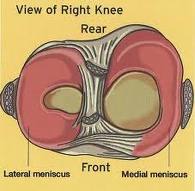
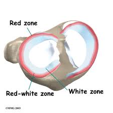
The meniscus cartilages in the knees are made of fibro-cartilage, similar to the end of the nose or ear. Meniscus type structures are found in many synovial joints. They provide cushion, stability and protection for the hyaline cartilage that is on the end of the bones of the knee joint. They are very resilient to compression (providing their cushioning function), but are susceptible to injury when torsion or twisting motions with pressure are applied. As we age, the blood supply (red zone below) to the meniscus diminishes, leaving these important protective cushions with minimal capacity to regenerate and heal after injury and use.
Traditionally, orthopedic surgeons felt that the meniscus was a vestigial–or unnecessary– structure that could be removed without detriment to the patient. After removal of entire meniscus cartilages in the 50s, 60s and 70s, it was soon realized that patients rapidly developed arthritis–or degradation–of the hyaline cartilage within 5-10 years. Thus, in the 70s and 80s, partial removal of the meniscus commenced, leaving some of the meniscus intact. This slowed the formation of arthritis from the 5-10 year range to the 10-15 year range. Arthroscopic techniques developed in the 80s and 90s led to the removal of smaller yet portion of the meniscus with less invasion of the knee joint.
In this series of posts, I will focus on the current regenerative and orthopedic care of meniscus injuries.