The meniscus is tough fibrocartilage between the thigh bone(femur) and shin (tibia). There are two meniscus per knee: one on the inside (medial) and one on the outside (lateral).
The meniscus functions as a shock absorber and therefore is critical.
The meniscus is C-shaped.
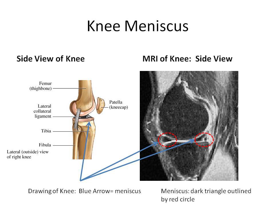
In viewing an MRI it is important to understand the view: frontal/side/ from the top or from the bottom.
When viewing the knee from the side, the meniscus are two dark triangles between the femur and tibia (outlined in red).
The meniscus is easily viewed from the front as demonstrated below.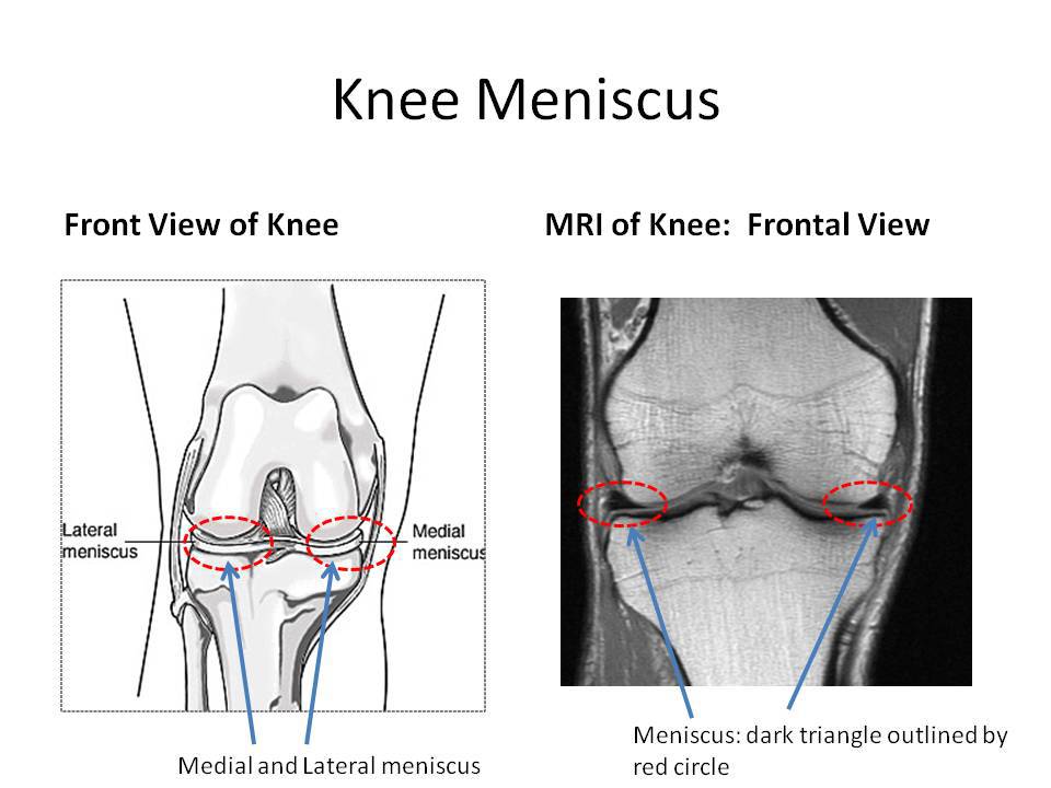
There are non-operative options to repair meniscus injuries which include the use of the stem cell therapy. The Regenexx procedure allow patients the opportunity to use their own mesenchymal stem cells to repair damaged knee tissue.